Download
1 / 58
680 likes | 1.27k Views
HEMOBLASTOSES. ANEMIAS. assistant of professor Nechiporenko G.V. HEMOBLASTOSES. HEMOBLASTOSES are tumoral diseases of hemo-lymphopoietic tissue. They are subdivided into: 1) Leukemias 2) Peripheral lymphomas.

E N D
HEMOBLASTOSES.ANEMIAS. assistant of professor Nechiporenko G.V.
HEMOBLASTOSES • HEMOBLASTOSESare tumoral diseases of hemo-lymphopoietic tissue. • They are subdivided into: 1) Leukemias 2) Peripheral lymphomas
Acute lymphoblastic leukemia (ALL) Lymphoblasts are very immature cells with large nuclei that contain nucleoli. ALL is more common in children than adults. Many cases of ALL in children respond well to treatment, and many are curable.
Chronic lymphocytic leukemia (CLL)Mature lymphocytes are increased markedly in number. A disease is most often seen in older adults;it responds poorly to treatment, but it is indolent.
Bone marrow of a patient with acute myeloblastic leukemia.There are many large immature myelocytes and one megakaryocyte at the right center
Blood smear, initial presentation of chronic myeloid leukemia (CML) - High power
Blood smears, chronic myelogenous leukemia (CML) blast phase (left)
The skull demonstrates the characteristic rounded "punched out" lesions of multiple myeloma.
Round lesions filled with a soft reddish material are indicative of foci of myeloma in this section of vertebral bone.
There are numerous plasma cells with eccentric nuclei and a perinuclear halo of clearer cytoplasm in a smear of bone marrow aspirate from a patient with multiple myeloma.
Here is a 5 cm lymph node (obviously from a patient with lymphadenopathy). The node should normally be soft and pink and less than 1 cm in size. This lymph node is involved with Hodgkin's disease.
This is a liver that is involved with Hodgkin's disease. The staging of Hodgkin's disease is very important in determining therapy.
Hodgkin's disease, nodular sclerosis type. Note the bands of pink collagenous tissue dividing the field in this lymph node.
These are the lacunar cellscharacteristic for the nodular sclerosis type of Hodgkin's disease.
This is a high power view of a Reed-Sternberg cell seen with Hodgkin's disease. Note the large, prominent nucleoli.
This is a malignant lymphoma, small cleaved cell type, follicular (also known as: malignant lymphoma, poorly differentiated lymphocytic type, nodular). • Here is a lymph node involved by lymphoma. The capsule of the node has been invaded and the lymphomatous cells extend into the surrounding adipose tissue. Note that the follicles are numerous and irregularly sized.
Malignant lymphoma is typically extranodal in AIDS. Seen here in small intestine are two mass lesions on the mucosal surface.
Extranodal malignant lymphoma inAIDS is often multifocal. Seen here in liver are two mass lesions on the cut surface. The color can range from white to tan to red, often intermixed.
Non-Hodgkin's lymphomas seen in the central nervous system with AIDS are essentially clonal expansions of Epstein-Barr virus infected lymphocytes. These lymphomas are high grade (immunoblastic) and agressive, with a poor prognosis.
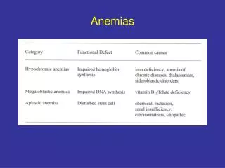
Anemia • Anemia is a reduction in the concentration of the hemoglobin in the blood. It is usually accompanied by reduction in the number of red blood cells (with the exception of iron-deficiency types, thalassemia). Poikilocytosis (different size), anisocytosis (different shape) of erythrocytes can develop in blood. Erythroblasts, normoblasts, megaloblasts appear also.
Classification of Anemias. A. PATHOPHYSIOLOGIC I. Anemia due to increased blood loss • Acute post-haemorrhagic anemia • Chronic blood loss II. Anemias due to increased red cell destruction (Hemolytic anemias) A. Extrinsic (extracorpuscular) red cell abnormalities B. Intrinsic (intracorpuscular) red cell abnormalities
III. Anemias due to impaired red cell production • a) Cytoplasmic maturation defects • Deficient haem synthesis:Iron deficiency anemia • Deficient globin synthesis:Thalassaemic syndromes • b) Nuclear maturation defects • Vitamin B12 and/or folic acid deficiency: Megaloblastic anaemia • c) Defect in stem cell proliferation and differentiation • Aplastic anemia • Pure red cell aplasia • Anemia of chronic disorders • Bone marrow infiltration • Congenital anemia