Download

1 / 27
300 likes | 513 Views
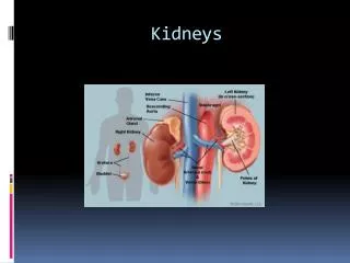
Cystic Diseases of Kidneys. Cystic renal diseases;. Cystic renal dysplasia (Potter II) Polycystic kidney disease 2.1. Adult (autosomal dominant) (Potter III) 2.2. Infantile (autosomal recessive) (Potter I) Medullary cystic disease 3.1. Medullary sponge kidney 3.2. Nephronophtisis

E N D
Cystic renal diseases; • Cystic renal dysplasia (Potter II) • Polycystic kidney disease 2.1. Adult (autosomal dominant) (Potter III) 2.2. Infantile (autosomal recessive) (Potter I) • Medullary cystic disease 3.1. Medullary sponge kidney 3.2. Nephronophtisis • Dialysis associated cystic disease • Simple renal cysts • Parenchymal renal cysts • Cystic Change with Obstruction(PotterIV) (Perihilar renal cysts)
1. Cystic renal dysplasia(Potter II) • The most common form of inherited cystic renal disease abnormal differentiation of the metanephric parenchyma during embryologic development of the kidney • Unilateral (affected person survivesin many cases) absence of one functional kidney from birth the other kidney undergoes compensatory hyperplasia may reach a size similar to the combined weight of two kidneys (400 to 500 g)
There are two main subgroups: Type IIa: affected kidney is large Type IIb: affected kidney is quite small (hypodysplasia) • Combinations: In unilateral cases: • one kidney or part of one kidney • Type II a or Type IIb In bilateralcases: • both can be large • both can be small • one can be larger and the other small
Gross: • The cysts - are variably sized (from 1 mm to 1 cm) - filled with clear fluid Microscopy: • Few recognizable glomeruli and tubuli(nephron) • The hallmark: presence of "primitive ducts" lined by cuboidal to columnar epithelium and surrounded by a collagenous stroma • Increased stroma may contain small islands of cartilage • The liver will not show congenital hepatic fibrosis • Differential diagnosis: Infantile (autosomal recessive) (Potter I)
2. Polycystic kidney disease 2.1. Adult (autosomal dominant): Potter III 2.2. Childhood (autosomal recessive): Potter I (1:30,000)
2.1. Autosomal dominant polycystic kidney disease(ADPKD); Adult polycystic kidney disease • Common (1:1,000) • Autosomal dominant pattern • High recurrence risk in affected families 50% • Rarely manifests itself before middle age - diagnosis by ultrasound - renal hypertension - progressive renal failure as the cysts become larger middle aged older adults • Half of these patients are on dialysis or transplanted
Pathology • Very large kidneys (3 or 4 kg or more) • Hundreds of fluid-filled cysts (up to 4 cm in diameter) • Hemorrhage into some cysts • The surrounding normal kidney tissue undergoes pressure atrophy • Surprisingly, they may still be working
ADPKDAssociated Conditions • Liver cysts (30%) • Splenic cysts (10%) • Pancreatic cysts (5%) • Cerebral aneurysms (20%) • Diverticulosis coli
Outstandig Clinical Findings: • high blood pressure • renal failure • intracranial hemorrhage (ruptured berry aneurysms) • cyst infections (nosocomial) • harmless hepatic, splenic and pancreatic cysts (US)
2.2. Autosomal recessive polycystic kidney disease(ARPKD); Infantile (Childhood) polycystic kidney disease • Autosomal recessive pattern • Bilateral • In utero poor renal function and failure to form significant amounts of fetal urine oligohydramnios + pulmonary hypoplasia so that newborns do not have sufficient lung capacity to survive
Huge, white, smooth-surfaced kidneys at birth • Cysts 1-2 mm in diameter (from the collecting ducts) • They are arranged in a radial, "sun-ray" pattern perpendicular to the capsule (because the collecting ducts are dilated) • Fatal in infancy or early childhood • Enormous kidneys restrict the ability of the lungs and gut to function • Differential diagnosis: Cystic renal dysplasia (Potter II) congenital portal fibrosis of the liver –present in • The liver will show congenital hepatic fibrosis • Differential diagnosis: Cystic renal dysplasia (Potter II)
Infantile type/autosomal recessive polycystic kidney disease (ARPKD).
3. Medullary cystic disease 3.1. Medullary sponge kidney 3.2. Nephronophtisis
3.1. Medullary sponge kidney • Idiopathic • Very common (1 in 200 people) • Normal renal functions • Dilated distal portions of collecting ducts superficially resemble cysts: Urinary stones within the "cysts“ Superimposed infection (pyelonephritis) • Chronic back pain
3.2. Nephronophthisis Uremic medullary cystic disease • Most common cause of endstage renal disease in children and young adults • Pathology: Cysts in the medulla Cortical tubular atrophy Interstitial fibrosis • The initial manifestations are inability to retain sodium and water
4. Dialysis associated cystic disease ("trans-stygian kidney") • Styx • A few remaining tubules stretched wide open ("cysts") stones painful bleeding aggressive carcinomas • Pathology fibrosis and a few chronic inflammatory cells oxalate crystals in the tubules fibromuscular masses in the blood vessels cortical adenomas and renal cell carcinomas
5. Simple renal cysts • A few cysts in a kidney • Old person • Commonest incidental finding at autopsy • Often develop after small kidney infarcts ("arterial nephrosclerosis")
6. Parenchymal renal cysts • 1. Associated with infection: TB Echinococcus • 2. Associated with tumor Cystic degeneration of a carcinoma • 3. Traumatic intrarenal hematoma
7. Cystic Change with Obstruction • PotterIV • Fetus and newborn with urinary tract obstruction renal cystic changes hydroureter hydronephrosis bladder dilation
Unilateral /Bilateral • depends upon the point of obstruction For example, posterior urethral valves in a male fetus, or urethral atresia in a male or female fetus, will cause bladder outlet obstruction so that both kidneys are involved
Pathology The cysts may be no more than 1 mm in size The cysts tend to be in a cortical location "cortical microcysts"