Download
1 / 44
500 likes | 555 Views
Chapter 6 The Genetics of Microorganisms. Structure of DNA. Two strands Nucleotides Hydrogen bonds between strands Neighboring deoxyribose connected 3’ of one deoxyribose to 5’ of next deoxyribose Phosphate in between Double helix Base pairing G and C A and T. Figure 6.1. Roles of DNA.

E N D
Structure of DNA • Two strands • Nucleotides • Hydrogen bonds between strands • Neighboring deoxyribose connected • 3’ of one deoxyribose to 5’ of next deoxyribose • Phosphate in between • Double helix • Base pairing • G and C • A and T Figure 6.1
Roles of DNA • Replication • cell division • need accurate copy • Gene expression • DNA • RNA • Protein Figure 6.2
DNA Replication • Semi-conservative • old strand-template • new strand-complementary • Replication fork • multiple enzymes • DNA unwinds • exposes nucleotides • synthesize new strand • one direction: 5’ to 3’ Figure 6.3
DNA Replication • Complementary nucleotides match (A=T; G=C) • DNA polymerase III binds nucleotides releasing pyrophosphate Figure 6.3
Bacterial chromosomes • Replication of circular chromosome • Origin of replication • bubble forms • DNA unwinds • Replication occurs in both directions • Two replication forks • Continues until replication forks meet • Strands separate Figure 6.4
Replication fork Figure 6.5
DNA Replication • Leading strand • replication is continuous (5’ to 3’) • primase makes primer • DNA added to primer • fork opens and replication continues • Lagging strand • polymerization in only one direction • can’t go 3’ to 5’ • short segments synthesized (Okazaki fragments) • when fork opens, new primer is made • synthesis in direction away from fork • fragments are joined together by DNA ligase
Transcription • RNA polymerase binds DNA at site of promoter Figure 6.6
Transcription • DNA unwinds • nucleotide bases are exposed Figure 6.6
Transcription • ribonucleotides pair with exposed bases • uracil in RNA replaces thymine • U binds A • ribonucleotides are polymerized into growing RNA chain Figure 6.6
Transcription • Termination sequence • Release of transcript • single strand RNA • DNA closes Figure 6.6
Transcription • Role of RNA from transcription • mRNA • template which encodes the protein • tRNA • transfer amino acids used to build the protein • rRNA • part of ribosome which is the site of protein synthesis • All used for translation
Translation • Production of proteins • Based on genetic information of DNA • Genetic code • Codon has three nucleotide • Four different nucleotides • 64 possible combinations • 20 amino acids • Redundancy • Nonsense codons Figure 6.8
Translation • tRNA • binds an amino acid • specific amino acid for each tRNA • Anticodon • recognizes codon • three nucleotide sequence in mRNA which encodes a specific amino acid • activated with ATP Figure 6.7
Translation • Ribosome binds to mRNA • specific region • start codon • Methionine • Ribosome binding region • Shine-Dalgarno sequence Figure 6.9
Translation • tRNA with appropriate anticodon and specific amino acid binds to the codon on the mRNA • A site • second tRNA binds in similar fashion • P site • two amino acids are joined in a peptide bond Figure 6.9
Translation • Ribosome moves along mRNA • first tRNA without amino acid is removed • second tRNA with both amino acids moves to P site Figure 6.9
Translation • New tRNA enters A site • Growing amino acid chain is transferred to new amino acid Figure 6.9
Translation • steps repeat • ribosome moves • one codon at a time • protein chain • one amino acid added for every codon Figure 6.9
Translation • Continues until nonsense (stop) codon is reached • no tRNA matches • ribosome is removed • protein chain is released Figure 6.9
Transcription and Translation • Simultaneous transcription and translation • mRNA chain is transcribed • translation begins • multiple ribosomes on single mRNA • polysome Figure 6.10
Transcription Production of regulatory proteins Bind DNA near the promoter Example: Lactose operon Interruption of transcription Attenuation Translation Ribosomal proteins Global regulation Catabolite repression Nitrogen regulation Phosphorus regulation Stringent response Heat shock proteins Regulation of genes
Transcriptional regulation lac operon • lacZ • lacY • lacA • regulated by lacI • Lactose absent • repressor binds • stops transcription Figure 6.11
Transcriptional regulation • Lactose present • repressor bound by product of lactose • allolactose • transcription occurs • gene products of all genes are made Figure 6.11
Attenuation • Histidine operon • Histidine present • Leader protein made • Translation occurring simultaneously with transcription • Requires histidine • Attenuator loop forms on mRNA • Displaces RNA polymerase • Stops transcription Figure 6.12a
Attenuation • Histidine absent • Leader protein not made • Not enough histidines to complete protein • Antiterminator loop forms • Prevents attenuator loop from forming • RNA polymerase continues Figure 6.12b
Regulation of translation • Expression of ribosomal proteins • Unused proteins bind to encoding mRNA • Inhibit translation Figure 6.13
Two component regulation • Phosphorylation of sensor • Phosphate passed to response regulator • Response regulator reacts with DNA changing gene expression • Increase • decrease Figure 6.14
Genetic Information • Genome • total DNA of a cell • most have single circular chromosome • some have linear chromosome • Plasmids • small, circular, extrachromosomal DNA • encode beneficial factors • resistance factors (antibiotic) • conjugative plasmids • transfer to other cells • Genotype: genetic makeup • Phenotype: appearance and function
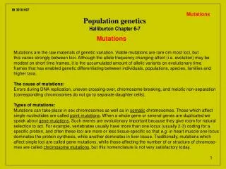
Mutations chemical change in DNA chemical mutagens Bind DNA Change in DNA physical mutagens UV light Ionizing radiation biological mutagens Transposable elements Insertion sequences transposons Changes in Genetic Information Figure 6.16
Types of mutations base substitution wrong nucleotide deletion mutation nucleotides deleted Inversion reverses order of a segment Transposition moves a segment of DNA Duplication identical new segment Results Lethal mutation Conditional expressed mutations Consequence of mutations Figure 6.15
Physical mutagen--UV damage • UV light • stimulates neighboring bases to form dimers • thymine dimers • activate repair systems Figure 6.17
Physical mutagen--UV damage • Thymine dimers distort the DNA structure • Enzymes remove the damaged nucleotides Figure 6.17
Physical mutagen--UV damage • Repairs may result in incorrect nucleotide replacement • Mutation is result Figure 6.17
Selecting and identifying mutants • Direct selection • Conditions favor growth of desired mutant • Growth of bacteria in presence of antibiotic • Only successful growth are mutants • Indirect selection • Prevent growth of mutant • Kill growing cells • Desired mutants larger percentage of population • Isolate mutants • Site-directed mutagenesis • Recombinant DNA manipulation
Selecting and identifying mutants • Brute strength • Screen large numbers • Replica plating • Transfer large numbers of colonies • Track growth Figure 6.18
Ames Test Figure 6.19
Transformation • DNA exits one cell, taken up by another cell • Natural • few bacteria take up DNA fragments • Artificial--induced in laboratory • useful tool for recombinant DNA technology Figure 6.20
Conjugation • Conjugative plasmids • plasmids transfer • genetically encoded • F plasmid in E. coli • sex pilus connect two cells • one cell F+ • one cell F- Figure 6.21
Conjugation • One strand of plasmid DNA is broken (nicked) • replication begins • synthesized linear strand enters F- cell • linear strand is copied forming a complete plasmid • both cells are F+ Figure 6.21
Transduction • Bacteriophage • virus that infects bacteria • reproduce in bacteria • some phages contain bacterial DNA • rare event • transducing particle • cell lysis and release • normal phage • transducing particles Figure 6.23
Transduction • Transducing particles • infect other bacteria • inject bacterial DNA into new cell • genetic exchange • one bacteria cell to another • integration into chromosome Figure 6.23
Eukaryotic Microorganisms • Genetic exchange • Similar to plants and animals • Haploid gametes fuse Figure 6.23