Download
1 / 40
400 likes | 409 Views
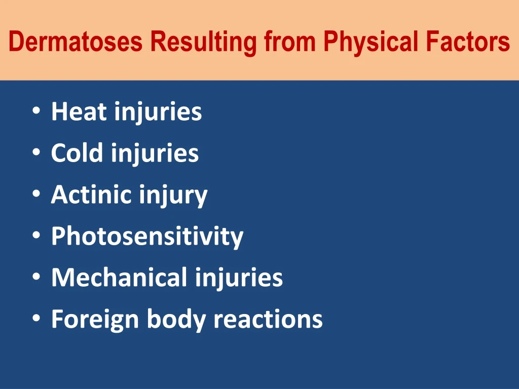
Dermatoses Resulting from Physical Factors. Heat injuries Cold injuries Actinic injury Photosensitivity Mechanical injuries Foreign body reactions. Heat injuries. 1.Miliaria

E N D
Dermatoses Resulting from Physical Factors • Heat injuries • Cold injuries • Actinic injury • Photosensitivity • Mechanical injuries • Foreign body reactions
Heat injuries • 1.Miliaria retention of sweat as a result of occlusion of eccrine sweat ducts, produces an eruption that is common in hot, humid climates The occlusion prevents normal secretion from the sweat glands, and eventually pressure causes rupture of the sweat gland or duct at different levels. escape of sweat into the adjacent tissue produces miliaria. several different forms are recognized :
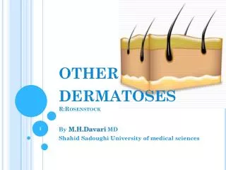
Miliariacrystallinasmall, clear, asymptomatic superficial vesicles with no inflammatory reaction. It appears in bedridden, bundled children. self-limited • Miliariarubradiscrete, pruritic, erythematouspapulovesiclesprickling or tingling. confluent on a bed of erythema. antecubitaland poplitealfossae,trunk, inframammary areas abdomen and inguinal regions • Miliariapustulosadermatitis produced injury of the sweat duct. pruritic superficial pustules, independent of the hair follicle, intertriginousareas (intertrigo), flexure surfaces of the extremities (Contact dermatitis), scrotum (lichen simplex chronicus).
Miliariacrystallina Miliariapustulosa Miliariarubra
MiliariaprofundaNon-pruritic, flesh-colored, deep-seated, whitish papules. asymptomatic, lasts only 1 h after overheating has ended, concentrated on the trunk and extremities.. • The most effective treatment for miliaria is to place the patient in a cool environment. Anhydrous lanolin and Hydrophilic ointment resolves the occlusion of pores and may help to restore normal sweat secretions. Mild cases may respond to dusting powders, such as cornstarch or baby talcum powder.
2.Erythema abigne • Persistent erythema or the coarsely reticulated residual pigmentation resulting from it that is usually produced by long exposure to excessive heat without the production of a burn. After the cause is removed, the affection tends to disappear gradually, but sometimes the pigmentation is permanent. • occurs on the legs as a result of habitually warming them in front of open fireplaces, space heaters, or car heaters, or the upper thighs with laptop computers. • Treatment with 5-fluorouracil (5-FU) or imiquimodcream. emollients containing α hydroxy acids or fluocinoloneacetonide 0.01 %, hydroquinone 4%, and tretinoin 0.05% may help pigmentation.
Cold injuries • 1.Acrocyanosis Acrocyanosis is a persistent blue discoloration of the entire hand or foot worsened by cold exposure. The hands and feet may be hyperhidrotic. • The cause is unknown. Smoking should be avoided. Acrocyanosisis distinguished from Raynaud syndrome by its persistent nature (as opposed to the episodic nature of Raynaud) and lack of tissue damage (ulceration, distal fingertip resorption). Patients with anorexia nervosa frequently manifest acrocyanosis as well as perniosis, livedoreticularis and acral coldness. It may improve with weight gain.
2.Chilblains (pernio) • localized erythema and swelling caused by exposure to cold. Blistering and ulcerations in severe cases. occur chiefly on the hands, feet, ears, and face • burning, itching, and redness call it to their attention. bluish-red lesions, the color partially or totally disappearing on pressure, and are cool to touch. • central cooling triggers peripheral vasoconstriction, keeping the whole body warm is critical. Smoking is strongly discouraged. • Nifedipine, 20 mg three times a day. Vasodilators such as nicotinamide, 500 mg three times a day,ordipyridamole, 25 mg three times a day, or sildenafil, 50 mg twice daily, may be used to improve circulation.
Actinic injury • 1.Sunburn and solar erythema • UV radiation (below 400 nm), visiblelight (400-760 nm), and infrared radiation (beyond 760 nm). Below 400 nm is the UV spectrum, divided into three bands: UV A, 320-400 nm; UVB, 280-320 nm; and UVC, 200-280 nm. • The minimal amount of a particular wavelength of light capable of inducing erythema on an individual's skin is called the minimal erythema dose (MED). UVB is up to 1000 times more erythemogenic than UVA. solar erythema is caused by UVB. Drug-induced photosensitivity, UVA is of major importance.
Sunburnis normal cutaneous reaction to sunlight in excess of an erythema dose. erythema 6 h after exposure followed by tenderness, in severe cases, blistering. chills, fever, nausea, tachycardia, and hypotension. Desquamation is common about a week after sunburn. • skin pigment changes: immediate pigment darkening within hours results from metabolic changes of melanin already in skin. Delayed tanning 2-3 days, and lasts 10-14 days,mediated through production of DNA damage.
Once redness and other symptoms are present, treatment of sunburn has limited efficacy. • Medium potency (class II) topical steroids applied 6 h after the exposure (when erythema first appears) give a small reduction in signs and symptoms. Therefore treatment of sunburn should be supportive, with pain management (using acetaminophen, Aspirin, or NSAIDs), plus soothing topical emollients or corticosteroid lotions.
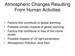
2.Ephelis (freckle) and lentigo • small less than 0.5 cm brown maculesoccur on the sun-exposed skin of the face, neck, shoulders, and backs of the hands. prominent during the summer and subside during winter. skin types I or II are susceptible. appear around age 5 . • lentigois a benign discrete hyperpigmentedmaculeat any age on any part of body. not dependent on sun exposure. • solar lentigoappears at a later age, persons with long-term sun exposure. backs of hands and face. • sun protection. Cryotherapy, topical retinoids, hydroquinone, lasers effective in solar lentigines.
freckles solar lentigo lentigo
3.Photoaging (dermatoheliosis) • Chronic sun exposure and chronologic aging are additive. Cigarette smoking important in development of wrinkles. the V area of the neck and chest, back and sides of the neck, face, backs of the hands and extensor arms, in women the skin between knees and ankles. • skin becomes atrophic, scaly, wrinkled, inelastic, with a yellow hue. Pigmentation is uneven, with a mixture of poorly demarcated hyperpigmented and white atrophic macules observed. The photodamaged skin appears generally darker because of these irregularities of pigmentation
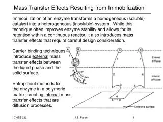
Poikiloderma of Civattereticulate hyperpigmentation with telangiectasia, and slight atrophy of the sides of the neck, lower anterior neck, and V of the chest. • Cutis rhomboidalisnuchaeskin on back of neck becomes thickened, tough, and leathery, and the normal skin markings are exaggerated. • Favre-Racouchot syndromeNodular elastoidosis with cysts and comedones occurs on the inferior periorbital and malar skin • Minimal trauma to the extensor arms leads to an ecchymosis, a phenomenon called actinic purpura. White stellatepseudoscars on the forearms are a frequent complication of enhanced skin fragility.
Cutis rhomboidalisnuchae Favre-Racouchot syndrome Poikiloderma of Civatte
Photosensitivity • Phototoxicity may occur from both externally applied (phyto-photodermatitis and berloque dermatitis) and internally administered chemicals (phototoxic drug reaction). • phototoxicity usually occurs on initial exposure, has an onset of less than 48 h, occurs in the majority of persons exposed to the phototoxic substance and sunlight, shows a histologic pattern similar to sunburn. • photoallergy occurs only in sensitized persons, have a delayed onset up to14 days (initial sensitization), histologic features of allergic contact dermatitis.
Phytophotodermatitis • Furocoumarinsin plants may cause a phototoxic reaction when come in contact with skin that is exposed to UVA light. • burning erythemafollowed by edema and development of vesicles or bullae. An intense residual hyperpigmentationmay persist for weeks or months. • Fragrance products containing bergapten or 5-methoxypsoralen is applied to the skin prior to exposure to sun berloquedermatitis may result. • cool compresses, mild analgesics and topical emollients. topical steroids and sun avoidance may protect against the hyperpigmentation which best managed by "tincture of time."
Idiopathic photosensitivity disorders • 1.Polymorphous light eruption • the most common form of photosensitivity. All races and skin types can be affected. The onset in the first four decades females outnumber males. The papular (or erythematopapular) variant is the most common, Scarring and atrophy do not occur. • lesions appear 1-4 days after exposure to sunlight. Areas of involvement face, the V area of the chest, neck, and arms. areas protected during the winter (extensor forearms) are particularly affected • appears most commonly in the spring. Often the eruption improves with continued sun exposure (hardening).
avoiding sun and high-SPF, broad-spectrum sunscreens. • topical tacrolimus ointment at night or twice daily, topical steroids,inseveral daily to weekly pulses, are necessary to control the pruritus and clear the eruption. • Antihistamines may be used for pruritus. • Systemic corticosteroids in short courses • hardening in the spring with UVB, narrow-band UVB, or psoralen + UVA (PUVA) • Systemic hydroxychloroquine sulfate, 200-400 mg/ day, may be used. • In severe cases azathioprine, cyclosporine, thalidomide, or mycophenolatemofetil may be considered.
2.Solar urticaria • most common in females aged 20-40 years. Within seconds to minutes after light exposure, typical urticarial lesions appear and resolve in 1-2 h, rarely lasting more than 24 h. • Chronically exposed sites may have some reduced sensitivity. In severe attacks, syncope, bronchospasm, and anaphylaxis may occur. • Broad-spectrum sunscreens should be used. Antihistamines, especially the non sedating H1 agents loratadine, cetirizine, and fexofenadine. PUVA or increasing UVA exposures
3.Chronic actinic dermatitis • Persistent, chronic, eczematous eruption in the absence of exposure to known photosensitizers. • affects middle-aged or elderly men. Skin lesions consist of edematous, scaling, thickened patches and plaques occur on exposed skin and may pare the upper eyelids, behind the ears, and the bottoms of wrinkles. In about one-third of patients, photopatchtesting yields a positive response • Therapy includes identifying possible topical photosensitizersMaximum sun avoidance and broad-spectrum sunscreens are essential. Topical tacrolimus is useful in some patients. Topical and systemic steroids are effective in some cases. Azathioprine, 50-200 mg/ day, is the most reproducibly effective treatment.
Mechanical injuries • 1.Callus • non penetrating, circumscribed hyperkeratosis produced by pressure. occurs on parts of body subject to intermittent pressure(palms and soles) and especially the bony prominences of the joints. • boxer's knuckle pads, tennis toe, prayer callus, neck callosities of violinists • callus has no penetrating central core,amore diffuse thickening. disappear spontaneously when pressure removed. • Ill-fitting shoes, orthopedic problems of the foot exerting abnormal pressure as in painful callosities of the feet. • Padding to relieve the pressure, paring of the thickened callus,useof such as 40% salicylic acid plasters.
2.Clavus (corns) • circumscribed, horny, conical thickenings with base on the surface and apex pointing inward • hard corns, which occur on dorsa of toes or on soles • soft corns between toes softened by macerating sweat. • In a hard corn, the surface is shiny and polished and, when the upper layers are shaved off, a core is noted in the densest part of the lesion. arise at sites of friction or pressure • bony spur or exostosisfrequently found beneath both hard and soft corns of long duration, and unless this exostosis is removed cure is unlikely. The soft interdigital corn usually occurs in the fourth interdigital space of the foot. These are soft, soggy, and macerated so that they appear white.
soft corns hard corns
Treatment by simple excision may be effective. • Plantar corns must be differentiated from plantar warts by paring off the surface keratin until either the pathognomonic elongated dermal papillae of the wart with its blood vessels, or the clear horny core of the corn can be clearly seen. • relief of pressure or friction by corrective foot wear • Soaking feet in hot water and paring the surface by means of a scalpel blade or pumice • 40% salicylic acid plaster is applied. After 48 h plaster removed, white macerated skin rubbed off
3.Pressure ulcers (decubitus) • pressure ulcer produced anywhere on the body by prolonged pressure. ischemia of underlying structures of skin, fat, muscles.complications include sepsis, local infection, osteomyelitis, fistulas. • lesion first appears as an area of persistent redness. Stage II is a superficial ulcer involving the epidermis and/ or dermis, stage III ulcers damaging the subcutaneous fat, and in stage IV, the muscle, bone, tendon, or joint capsule. • Prevention : redistributing pressure at an interval of 2 h. • frequent change of position, air-filled products, management of bacterial colonization, adequate nutrition. Debridement. Normal saline better than peroxide or povidone-iodine. anaerobic organisms colonize ulcers and cause a putrid odor. topical metronidazole eliminates odor.
4.Friction blisters • vesicles or bullae may occur at sites of combined pressure and friction,enhancedby heat ± moisture. • The feet of military recruits in training and the fingers of drummers ("drummer's digits") • If the skin is tense and uncomfortable, the blisters should be drained, but the roof should not be completely removed as it act as its own dressing. • pretreatment with a 20% solution of aluminum chloride hexahydrate for at least 3 days has been shown to reduce foot blisters significantly
Foreign body reactions • 1.Tattoo • introduction of insoluble pigments into the skin. Pigment is applied to skin and then needles pierce the skin • Pigments utilized may be carmine, India ink, chrome green, magnesium (lilac color), Venetian red, titanium (white color) • Tattoo-associated dermopathies may be reactive (allergic, lichenoid, granulomatous, or photosensitive) or infective (syphilis,hepatitis, tuberculosis, HIV, warts, molluscum and Hansen's disease), or may induce a Koebner response in patients with active lichen planusor psoriasis. • topical or intralesional steroids. Excision when lesions are small enough. Q-switched 532 nm neodymium:YAG laser.