Download
1 / 129
1.37k likes | 1.8k Views
THE MANAGEMENT OF ACUTE PANCREATITIS. Professor Ravi Kant MB, MS, FRCS (Edinburgh), FRCS (Glasgow), FACS, FICS, DNB, FAIS, FAMS,. INTRODUCTION. Introduction. “In the growing world of EBM, only 30% of surgery is based on evidence while 70% of medicine is evidence based” EJS, Sep 2005.

E N D
THE MANAGEMENT OF ACUTE PANCREATITIS Professor Ravi Kant MB, MS, FRCS (Edinburgh), FRCS (Glasgow), FACS, FICS, DNB, FAIS, FAMS,
Introduction • “In the growing world of EBM, only 30% of surgery is based on evidence while 70% of medicine is evidence based” EJS, Sep 2005
Introduction • Stress will be on Diagnosis, workup, prognostic predictors and management • Basic sciences
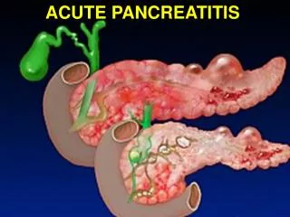
Definition • “Acute pancreatitis”: • Inflammation of the pancreas without, or with minimal fibrosis.
Epidemiology • 300,000 annually in US • 10-20% are severe • Total annual cost of 2 billion $$$ • (Biliary + alcoholic) 90% • Even in the west, biliary pancreatitis is the most prevalent type. • Incidence among AIDS patients 4-22%
Epidemiology • Local statistics?
Epidemiology • “Profile of acute pancreatitis in Jizan, Saudi Arabia” Saudi Med J. 2003 Jan;24(1):72-5. • (KFCH), Jizan, KSA over 12 years regional • 42% (biliary), 18% Post ERCP • “Pattern of acute pancreatitis” Saudi Med J. 2001 Mar;22(3):215-8. • Cross sectional, 2 years, Asir central hospital • 68% found to be biliary
Causes Biliary tract disease Alcohol Hyperlipedemia Hypercalcemia Trauma ERCP Ischemia Pancreatic neoplasia Pancreas divisum Ampullary lesions Duodenal lesions Infections Venom Drugs idiopathic Pathophysiology
Pathophysiology • Theories behind mechanism of biliary pancreatitis • Common channel theory • Incompetent sphincter theory • Co-localization theory
PATHOPHYSIOLOGY • Common channel theory • “Opie 1901” • Detergent effect of bile
Pathophysiology • Critique of common channel theory • Higher hydrostatic pressure in PD • Introduction of bile into PD in animal models failed to cause AP
Pathophysiology • Incompetent sphincter theory • Incompetent sphincter of Oddi due to stone passage reflux AP • Critique • How come papillotomy doesn’t routinely cause AP??
Pathophysiology • Co-localization theory “Steer & Saluja” 1998 • Most acceptable • Stones PD ductal hypertension ducutle rupture • Ductal pH = 9 …… parynchemal pH = 7 • trypsinogen + cathepsin B trypsin autodigestion cascade
Pathophysiology • Support of co-localization theory • CA-074me (cathepsin B inhibitor) prevented AP in 2 different models of acute pancreatitis
Pathophysiology • Alcoholic pancreatitis • No such thing as acute alcoholic pancreatitis • It is actually the first attack of chronic alcoholic pancreatitis
Diagnosis • Clinical picture • Investigations • “Acute pancreatitis is a diagnosis of exclusion” Schwartz’s
Diagnosis • Hx: • Epigastric pain • Radiating to back • Nausea, vomitting • Precipitating factor?
Diagnosis • Physical • V/S variable • Epigastric tenderness • Cullen’s / Grey Turner’s (1%) • Findings of complication(s)
Diagnosis • Serum markers • Amylase • Easiest to measure and most widely used • Rises immediately • Peaks in few hours • Remains for 3-5 days • “Three fold rise is diagnostic” • May be normal in severe attacks • May be falsely negative in hyperlipedimic patients • Inverse correlation between severity and serum amylase level • No need to repeat
Diagnosis • Serum markers • Urine amylase • Remains elevated for a few more days • Increase excretion of amylase with attacks of AP • Of great value when dealing with severe pancreatitis
Diagnosis • Serum markers • P/S – amylase • P amylase increases specificity to 93% • Lipase • “the serum marker of highest probability of disease” • Specificity of 96% • Remains elevated for longer time than total amylase
Pancreatitis p Choledocolethiasis p Parotitis s Renal failure s/p Liver cirrhosis s/p perforated bowel p mesenteric infarction p intestinal obstruction p Appendicitis p Peritonitis. P Gyne disease s Malignancies Lung CA Ovarian CA pancreatic CA Colonic CA pheochromocytoma; Thymoma multiple myeloma breast cancer Causes of hyperamylesemia
Radiology • Diagnostic role • X-ray • U/S • CE-CT
Radiology • X-ray • Air in the duodenal C loop • Sentinel loop sign • Colon cutoff sign • All these signs are non specific
Radiology • CE-CT • Enlargement of the pancreas • (focal/diffuse) • Irregular enhancement • Shaggy Pancreatic contour • Thickening of fascial planes • fluid collections. • Intraperitoneal / retroperitoneal • Retroperitoneal air
Radiology • U/S • Diagnosis of gallstones • F/U of pseudocysts. • Dx pseudoaneurysms • EAUS vs. EUS
Prognosis • Course either mild or severe • Mild = edematous pancreatitis • Severe = necrotic pancreatitis • No such thing as moderate pancreatitis
Prognosis • Serum markers • CT • Systemic complications • Prognostic scores • Ranson • Apache II • Modified Glasgow • Atlanta Atlanta Consensus 1992
Prognostic scores • Ranson’s • Published in 1974 • Predictor of morbidity/mortality • <2 0% mortality • 3-5 10-20% • >7 >50% mortality • Critique of Ranson’s • 11 parameters • 48 hours • No predictor value beyond 48hrs • Too pessimistic for today’s healthcare system
Prognostic scores • APACHE II • Immediate • Acute and chronic parameters • Complicated • >7 = severe pancreatitis
Prognostic biochemical markers • Biochemical markers of prognosis • Ideally • High sensitivity • High specificity • Discriminate severe from mild • Immediate • Widely available • Amylase & lipase • Highly sens./spec. • Lack prognostic value
Prognostic biochemical markers • Alternatives • CRP • 2 macroglobulin • PMN elastase • 1 antitrypsin • Phospholipase A2 • “CRP seems to be the marker of choice in these settings” • CRP >150 is diagnostic of severe pancreatitis
Prognostic biochemical markers • Other markers • IL-6 • Urinary TAP • These showed great promise in models and clinical trials • Failed in larger scale trials
CT scan (prognostic aspect) • “CT scanning with bolus IV contrast has become the gold standard for detecting and assessing the severity of pancreatitis” • “Currently, IV bolus contrast enhanced CT scanning is routinely performed on patients who are suspected of harboring severe pancreatitis, regardless of their Ranson’s or APACHE scores” Schwartz’s