Download

1 / 40
470 likes | 1.04k Views
Hemorrhagic diatheses in children. Sakharova I. Ye., M.D, Ph.D. The stopping of a bleeding is carried out due to three parts of a hemostasis:. Vascular integrity. Qualitative and quantative characteristics of platelets. Presence of coagulation factors in blood.

E N D
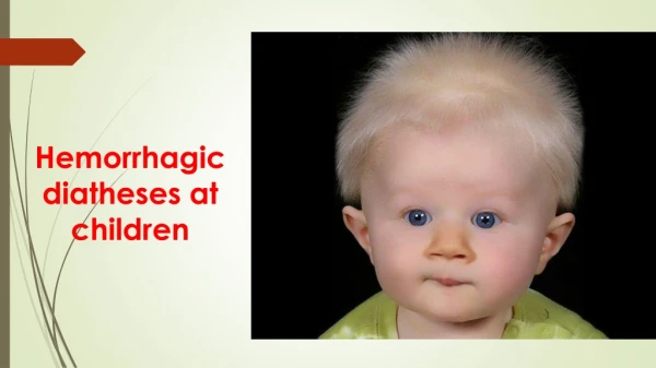
Hemorrhagic diatheses in children Sakharova I. Ye., M.D,Ph.D
The stopping of a bleeding is carried out due to three parts of a hemostasis: • Vascular integrity. • Qualitative and quantative characteristics of platelets. • Presence of coagulation factors in blood.
According to this all hemorrhagic diatheses are divided into 3 groups:1.Vasopathies 2.Thrombopathias 3.Coagulopathies
Schönlein-Henoch purpura(synonims - anaphylactoid purpura, allergic angiitis, small-vessel vasculitis, hemorrhagic vasculitis, Henoch-Schönlein disease) is one of the collagen vascular diseases in which basis lays immune complex mechanism of small vessels wall damaging with skin, joints, intestine and kidneys affection.
Clinical features. • The skin rush: urticarial initially then fades, to be replaced by symmetrical macular or papular- macular hemorrhagic purpuric lesions. They may remain small and discrete or enlarge and become quite blotchy, at times confluent. Sometimes in the center of blots can be necrosis. • Typical places of rush localization: extensor surfaces of legs, on the feet, over the joints, on the buttocks; occasionally, they may occur on the hands, extensor surfaces of arms, elbows, and face, but very rarely on the trunk.
Clinical features of the Schönlein-Henoch purpura. • joints involvement • acute abdominal pain, vomiting, melena • renal involvements (microscopic hematuria, with or without proteinuria) • scrotal involvement (epydidimitis, orchitis, and scrotal bleeding)
Laboratory findings: • General blood count reveals normochromic anemia, eosinophilia. • Mild leukocytosis and elevated ESR, which are associated with inflammatory process. • The platelet count, platelet function test, and bleeding time are normal. • Blood coagulation studies are normal. • Urinalysis frequency reveals hematuria, proteinuria.
Laboratory findings: • The ASO (antistreptolizin-O) titer is frequently elevated and the throat culture positive for group A beta-hemolytic streptococcus. • Serum Ig A may be elevated. • Circulating immune complexes are commonly present. • Positive C-reactive protein, increased level of sialic acids (acute phase reactants). • Hypercoagulation orientation of hemostasis parameters.
Basic therapy of hemorrhagic vasculitis: Antiaggregants (disaggregants) for 3-4 weeks Kurantil (Dipiridamol) 2-4 mg/kg/day (IV, IM, per os) Trental 5-10 mg/kg/day (IV, per os) Tiklopedin (Tiklid) 250 mg 3 times /day Direct anticoagulants for 3-4 weeks (under PTT control) Heparin 200-300 U/kg/day (IV, SC) Fracsiparin (Calciparin, Enocsiparin) 5000-7500 U/day (SC)
Basic therapy of hemorrhagic vasculitis: Fibrinolysis activators - Ac. nicotinici (IV) Nonsteroidal antiinflammatory drugs, NSAID for 2-3 weeks Aspirin 5-10 mg/kg 1 time in morning Indomethacin 2-4 mg/kg/day Ortofen 1-2 mg/kg/day
Other treatment: • Corticosteroids: prednisone 3-4 mg/kg/day for 5-7 days (corticosteroids therapy may provide symptomatic relief for severe gastrointestinal or joint manifestations, but doesn’t alter skin or renal manifestations) • Cytostatics (methotrexate – 50 mg/m2, azathioprine – 50-75 mg/day or cyclophosphamide 100-150 mg/day) • Penicillin in full therapeutic doses for 10 days (if culture for group A beta-hemolytic streptococcus is positive or if the ASO titer is elevated) • Antihistamines and less sedating agents (in patients with urticarial lesions) • Plasmapheresis with substitution of 2-5 plasma volumes
Idiopathic thrombocytopenic purpura (ITP, primary immune thrombocytopenic purpura, autoimmune thrombocytopenic purpura) describes an autoimmune disorder in which the number of circulating platelets is less than 150 G/l.
Clinical features. • The onset of the disease is usually sudden. The symptoms of intoxication and fever usually are absent. • Skin purpura, which arises either spontaneously or secondary to trauma. The type of rush is petechial-bruise. Petechiae may be found anywhere over the skin. Ecchymoses are usually found on the anterior surfaces of the lower extremities, over bony prominences such as the ribs, scapula, shoulders, and legs. Petechiae may be found in the conjunctiva, oral cavity mucose, in the soft palate. It is significant that rush is polymorphous and polychromatic.
Clinical features. • The second frequent clinical sign are bleedings. In the beginning of the disease can be nose bleeding (epistaxis), bleeding from gums, mucous membranes, gastrointestinal tract, kidneys and metrorrhagias (uterinal bleedings). Hemorrhage into the central nervous system, the most serious complication of thrombocytopenia.
Laboratory findings: • A marked decrease or absence of platelets • Prolonged bleeding time by Duke (> 4 min) • Poor clot retraction (normally occurs within an hour at 37 ºC) • Bone marrow examination often the increased number of immature megacaryocytes with a markedly basophilic cytoplasm • Antiplatelet antibodies are present in blood serum • Abnormal tourniquet test
Children who have platelet counts >30,000/mm3(30 x 109 /l) and are asymptomatic or have only minor purpura do not require routine treatment. Children who have platelet counts <20,000/mm3(20 x 109 /l) and significant mucous membrane bleeding and those who have platelet counts <10,000/mm3(10 x 109 /l) and minor purpura should receive routine treatment.
Treatment of ITPI stage (acute heteroimmune ITP) • Prednisolon 1 mg/kg/day • Dicynon 0.25 mg x 3 times/day or 12,5 % 1-3 ml; Doxium, Androkson • Vitamin C, K, calcium medicines • Antifibrinolytic agents:aminocaproic acid 0,05-0,1 g/kg/day
Treatment of ITPII stage (chronic autoimmune ITP) • Prednisolon 1 mg/kg/day • Intravenous immune globulin (IVIG) 400 mg/kg/dayduring 2-5 days – IG “Biochemi” Austria, endoglobulin, Austria; sandoglobulin, Switzerland; intraglobulin, Germany • Anti-D(Rh) immunoglobulin (intravenous Rh immune globulin) • ά2-interferon • Plasmapheresis
Treatment of ITPIII stage • Splenectomy IV stage • Steroid use and immunosuppressives therapy According to a recent study, using a combination of weekly vincristine, weekly methylprednisolone, both until platelet counts reached 50,000/mm3, and cyclosporine orally twice daily until the platelet count is normal for 3-6 months seems promising, though larger prospective studies are needed.
Wiskott-Aldrich syndrome(congenital thrombocytopenia) – an X-linked disorder, the initial manifestations are often present at birth and consist of petechiae, bruises and bloody diarrhea due to thrombocytopenia. The classic triad of this syndrome includes thrombocytopenia, eczema and immunodeficiency
Von Willebrand's disease(vWD)is inherited as an autosomal dominant disease. There is deficient or defective production of von Willebrand factor. This protein mediates platelet adhesion to the endothelium and protects factor VIII from degradation.
Can be classified as Type I Type II Type III pseudo-vWD, based on their clinical history and laboratory evaluation
Laboratory findings: • The platelet count is normal • Prolonged protrombin time (normal 12-15 sec.) • Prolonged bleeding time by Duke (> 4 min) • Decreased ristocetin cofactor activity in plasma • Low level of antihemophilic globulin (AHG – F VIII) • Reduced platelets adhesiveness
Treatment Depends from the type of the disease: Type I - Desmopressin acetate (DDAVP), an analogue of vasopressin 0,3 g/kg IV or intranasal DDAVP (Stimate) Type II, III – cryoprecipitate or platelet transfusions
Bernard-Soulier syndromeis an autosomal recessive disorder with characteristic easy bruizability and severe bleeding in injury. Lab. Findings: • the platelet count is normal or slightly decreased • bleeding time is prolonged • platelets are very large in peripheral blood smear.
Hemophilia A and Bare inherited bleeding disorders caused by deficiencies of clotting factor VIII (F VIII - antigemophilic globulin (AHG)) and factor IX (F IX - plasma thromboplastin component (PTC) or Christmas factor) correspondingly.
xx Xy Xy Xy Xx Xx xy xy xy xx Xx xy XX Xx Xy xy Mechanism of hemophilia inheritance
Laboratory diagnostic of hemophilia • Prolongedcoagulation time (normal 5-10 minby Lee-White) • Prolonged recalcification time (normal 80-140 sec) • Prolonged heparin time (normal 11-16 min) • Prolonged protrombin consumption (partial thromboplastin) time (normal25-39sec)
Prophylactic treatment The aim of this treatment is to maintain 5 % factor activity in patient’s blood. Start from the age of 1-2 years. Use monoclonal-antibody purified FVІІІ , F ІХand recombinant FVІІІ , F ІХ 3 timesin weekin hemophilia Aand 2 times in week in hemophilia B 25-40 IU/kg.
Treatment of acute bleeding episodes Hemophilia A • Fresh frozen plasma(100 ml=80IU АHG) Dose is 10-15 ml/kgIVduring 30-60 min, repeat after 8-12 hours. • Cryoprecipitate – Monoclonal-antibody purified FVІІІand recombinant FVІІІ Hemophilia A – Fresh frozen plasma – Monoclonal-antibody purified FIX and recombinant FIX
During severe or dangerous (e.g. CNS, retroperitoneal) bleeds need to obtain 50-100% factor activity for 7-10 days. For less critical situations (e.g. dental extractions, haematuria, soft tissue bleeds), 20-50% factor activity for 2-7 days are generally sufficient. For uncomplicated haemarthroses or superficial muscle or soft tissue bleeds, 20-30% for 1-2 days.
Laboratory differential diagnostics of hemorrhagic diatheses Count of platelets Time of bleeding Coagulation time Clot retrac-tion Coagulogram Hem. vaskul. ITP Trombopathia Hemophilia n n n n n n n n n n n n n n n n