Download
1 / 55
601 likes | 1.46k Views
Update in nephrology Contrast induced nephropathy, nephrogenic systemic fibrosis and acute phosphate nephropathy. Steven D. Weisbord MD, MSc , FASN Renal-Electrolyte Division University of Pittsburgh School of Medicine.

E N D
Update in nephrology Contrast induced nephropathy, nephrogenic systemic fibrosis and acute phosphate nephropathy Steven D. Weisbord MD, MSc, FASN Renal-Electrolyte Division University of Pittsburgh School of Medicine
1) A 45 year old WM with a serum creatinine of 1.0 mg/dL is undergoing a procedure. He is at risk for: • Contrast induced nephropathy • Acute phosphate nephropathy • Nephrogenic systemic fibrosis • Contrast nephropathy and acute phosphate nephropathy • Contrast nephropathy and nephrogenic systemic fibrosis • Acute phosphate nephropathy and nephrogenic systemic fibrosis • All of the above
2) A 45 year old WM with a serum creatinine of 1.8 mg/dL is undergoing a procedure. He is at risk for: • Contrast nephropathy • Acute phosphate nephropathy • Nephrogenic systemic fibrosis • Contrast nephropathy and acute phosphate nephropathy • Contrast nephropathy and nephrogenic systemic fibrosis • Acute phosphate nephropathy and nephrogenic systemic fibrosis • All of the above
3) A 45 year old WM on chronic hemodialysis is undergoing a procedure. He is at risk for: • Contrast nephropathy • Acute phosphate nephropathy • Nephrogenic systemic fibrosis • Contrast nephropathy and acute phosphate nephropathy • Contrast nephropathy and nephrogenic systemic fibrosis • Acute phosphate nephropathy and nephrogenic systemic fibrosis • All of the above
Pathophysiology of CIN Radiocontrast Administration Intrarenal Vasoconstriction Rheologic Effects Direct Cytotoxicity Osmotic Load Generation of ROS Medullary Hypoxia CIN
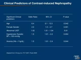
Risk factors for CIN • Patient-related • Renal insufficiency • Diabetes mellitus* • Intravascular volume depletion • Reduced cardiac output • Concomitant nephrotoxins • Procedure-related • ↑ volume of radiocontrast • Multiple procedures w/i 72 hours • Intra-arterial administration • Type of radiocontrast } additive risk * Diabetes alone not strong risk factor
Renal Insufficiency and Diabetes Mellitus McCullough PA et al. Am J Med. 1997;103:368-375.
Approach to screening with SCr • Known renal insufficiency • Diabetes mellitus • Proteinuria • Advanced age • Hypertension • Nephrotoxic drug use • History of kidney problem after radiocontrast • Advanced liver disease Consider screening SCr if pt has 1 or more of these: Weisbord SD, my approach
Relationship Between Serum Creatinine and eGFR 59 ml/min/1.73m2 36 ml/min/1.73m2
Implications of CIN • CIN may result in any or all of the following: • Delay in discharge of patient • Permanent kidney damage • Dialysis • Increased patient mortality Dangas G et al. Am J Cardiol. 2005;95:13-19.
CIN and mortality Adjusted OR: 5.5; p<0.01 Levy et al. JAMA 1996; 275:1489-1494
CCB Loop diuretics* Mannitol* Dopamine* Fenoldopam* ANP Hemodialysis* NAC Theophylline Aminophylline Ascorbic acid Statins Hemofiltration Preventive strategies for CIN Ineffective Unclear benefit Effective • IVF • Choice of contrast * Possibly harmful
NAC for CIAKI (n=83) 21% % CIN (Scr ↑ 0.5 mg/dL @ 48h) P=0.01 2% NAC Control Tepel M, et al. N Engl J Med 2000; 343:180-184
Meta-Analyses of NAC ** Given degree of heterogeneity, calculation of summary estimate would be invalid
NAC - summary • Protective effect unclear • Many studies to date have methodological flaws • Cheap and benign (in oral form) • Should not be used in lieu of other measures
Clinical trials of volume expansion • 1994 → present • Provide clinical basis for: • Protective effect of IVF • Deleterious effect of furosemide • Superiority of isotonic IVF • Superiority of IVF to pt-directed oral fluids • Potential benefit of oral NaCl
Rate of CIN: 11% 28% 40% Solomon R, Werner C, Mann D, D’Elia J, Silva P. N Engl J Med. 1994;331:1416-1420.
Isotonic v. hypotonic saline P=0.04 P=0.93 P=0.35 Mueller C, et al. Arch Int Med. 2002; 162:329-336
Saline vs. Bicarbonate IV fluid (8/59) P = 0.02 (1/60) Merten et al. JAMA 2004;291:2328-2334
Meta-analysis of NaCl v. NaHCO3 OR 0.46 [0.26-0.82] Navaneethan SD et al. 617-627; 2009; American Journal of Kidney Diseases
IV NaCl v. oral NaCl P=NS % CIN N=76 N=79 N=77 N=80 Dussol et al. Nephrology Dialysis Transplantation. 2006;21:2120-2126
Meta-analysis of IOCM v. LOCM P=0.003 IOCM LOCM NS NS % pts NS McCullough et al. JACC 2006;48:692-9
Meta-analysis of IOCM v. LOCM Favors IOCM Favors LOCM Heinrich et al. Radiology 2009;250:68-86
Summary of prevention • NAC – of unclear benefit • I use 1200 mg po bid x 2 days • IV fluids beneficial – isotonic >> hypotonic • ? Superiority of NaHCO3 • Abbreviated regimen OK – 1 hr pre and 4-6 hr post • Low or iso-osmolal contrast • Mixed data on superiority of iso-osmolal
Summary of CIN • Remains common due to high use of iodinated contrast • Risk factors well known – CKD • Adverse outcomes with CIN • Prevention: • Isotonic IV fluids • NAC - ? benefit • Choice of contrast
NSF - History and Nomenclature • Disease initially identified in late 1990s as fibrosing skin condition • Named nephrogenic fibrosing dermopathy (NFD) • Subsequently found to also have systemic manifestations • skeletal muscle, lung, liver, testes, myocardium • most prominent findings are dermatologic • Re-named Nephrogenic Systemic Fibrosis (NSF) Cowper SE. Available at http://www.icnfdr.org; Deo A et al. Clin J Am Soc Nephrol. 2007;2:264-267.
NSF: Skin manifestations • Distribution • Usually symmetrical • Extremities trunk • Face/neck typically spared • Signs • Swelling and erythema of extremities • Induration: distal proximal • Woody papules • Symptoms • Burning, itching, pain • Reduced flexibility immobility • Muscle weakness } Can be very disabling Marckmann P et al. Clin Nephrol. 2008;69:161-168; Mitka M. JAMA. 2007;297:252-253; Thomsen HS. Eur Radiol. 2006:16:2619-2621;Cowper SE. http://www.icnfdr.org.Issa N et al. Cleve Clin J Med. 2008;75:95-111
NSF: Epidemiology • No gender predilection • Affects patients of all ages; most commonly middle age • Affects various ethnic/racial groups • Seen in North America, Europe, and Asia • Only seen in pts with kidney disease Cowper SE. Available at http://www.icnfdr.org.
NSF: Clinical appearance Occurs days to many months after exposure to GBCA Marckmann P et al. Clin Nephrol. 2008;69:161-168
Stage I Stage II Stage III Stage IV Stage V 130 120 110 100 90 80 70 60 50 40 30 25 20 15 10 5 0 GFR NSF: Association With Renal Disease • All reported NSF in pts with renal impairment • Reported in stages 4-5 CKD • eGFR <30 mL/min/1.73 m2 • Most commonly dialysis pts Most cases NSF Issa N et al. Cleve Clin J Med. 2008;75:95-111.
Markmann et al – 13 of 370 ESRD pts (3.5%) Deo et al – 3 of 87 ESRD pts – (3.4%) Broome et al – 12 of 301 HD-pt exposures (4%) Prince et al – 1 of 265 ESRD pt (0.4%) Incidence after GBCA in ESRD = 0.4-4% based on retrospective analyses NSF: Incidence After GBCA in End-Stage Renal Disease Markmann P et al. J Am Soc Nephrol. 2006:2359-62; Deo A et al. Clin J Am Soc Nephrol. 2007:264-7; Broome DR et al. AJR 2007:586-92; Prince MR et al. Radiology. 2008;248:807-816.
Incidence of NSF in stages 4 and 5 CKD following MRA • 2 retrospective analyses of: • Large tertiary referral center in UK • Database of pts screened/enrolled in ASTRAL study • Results: • 0 of 252 pts with eGFR < 30 developed NSF • 0 of 485 pts with eGFR < 60 developed NSF • 1 of 1735 pts (0.06%) with CKD developed NSF (data extrapoloated and pt with NSF had stage 4-5 CKD) Chrysochou et al. Journal of Mag Reson Imag 29:887-94
Gd is a lanthanide ion Free Gd is highly toxic Contrast agents for MR imaging – metal ion (Gd) bound to ligand (Gd-chelate complex) GBCA – excreted by the kidneys With impaired kidney function, T1/2 of GBCA increases ? displacement of Gd from chelate (transmetallation) Tissue exposure of Gd ? Fibrosis leading to NSF NSF – Pathogenesis and GBCA Perazella MA. Clin J Am Soc Nephrol. 2007;2:200-202; Rofsky NM et al. Radiology. 2008:608-12.
NSF: Speculative Pathogenesis 1 4 5 2 3 cF, circulating fibrocyte; Cyto, cytokinesPerazella MA. Clin J Am Soc Nephrol. 2007;2:200-202.
NSF and Specific GBCA Volunteer case reports to MedWatch (FDA) as of 10/07 Penfield JG et al. Semin Dial. 2008;21:129-134.
Risk factors for NSF - GBCA - GBCA strongly associated with NSF - Few case reports of NSF without known GBCA exposure Agarwal R et al. 2008 Nephrol Dial Transplant: 1-7; Wahba M. et al. Amer J. Transplant 2007;7:2425-32
NSF and dose of GBCA • Retrospective review - biopsy-confirmed NSF cases from 1997–2007 in 2 large hospitals • 83,121 pts received GBCA • 15 cases of NSF confirmed after GBCA • 74,124 pts - low dose GBCA (0.1 mmol/kg) – NSF = 0% • 8,997 pts – high dose GBCA (0.2-0.4 mmol/kg) – NSF = 0.17% Prince MR et al. Radiology. 2008;248:807-816.
NSF and pro-inflammatory state Sadowski EA et al. Radiology. 2007;243:148-157.
Hypercoagulability states Surgical procedures Esp, reconstructive vascular components Hepatic disease Hepatorenal syndrome Liver transplantation Hepatitis B and C Thrombotic events Idiopathic pulmonary fibrosis SLE Hypothyroidism serum Ca or PO4 Hyperparathyroidism Metabolic acidosis NSF: Associated Clinical Conditions • These conditions may be associated with increased use of MRI • Some conditions result from or cause renal disease Issa N et al. Cleve Clin J Med. 2008;75:95-111.
NSF and other forms of renal disease • Acute kidney injury appears to be a risk factor for NSF • Acute kidney injury an “inpatient” disease • No need to routinely screen outpatients for acute kidney injury before MR • Peritoneal dialysis (PD) • Appears to be risk factor - ? > HD • Reduced clearance of Gd with PD Joffe P et al. Acad Radiol. 1998;5:491-502; Prince MR et al. Radiology. 2008;248:807-816.
Oral steroids (eg, prednisone) Topical Dovonex (under occlusion) Extracorporeal photopheresis Plasmapheresis Cytoxan Thalidomide Ultraviolet therapy Physical therapy Pentoxifylline High-dose IV Ig therapy Renal transplantation IV sodium thiosulfate NSF: attempted treatment strategies - Most evidence anecdotal and/or unconfirmed. - Improving renal function may slow, arrest, and reverse NSF - PREVENTION IS KEY !!!! Cowper SE. Available at http://www.icnfdr.org; Issa N et al. Cleve Clin J Med. 2008;75:95-111.
Prevention: FDA recommendations on use of GBCA • Screen all pts for renal dysfunction: history and/or lab tests • Avoid GBCA in pts with known risks for NSF unless diagnostic information cannot be obtained with non-contrast MR or other diagnostic procedures • When administering GBCA: • Do not exceed recommended GBCA dose in product labeling • Allow sufficient time for elimination of GBCA from the body prior to any re-administration • For pts on HD, consider prompt HD following GBCA http://www.fda.gov/cder/drug/InfoSheets/HCP/gcca_200705.htm
Summary of NSF • Debilitating fibrosing condition - 10 skin findings • Associated with gadolinium contrast agents • Risk factors – high dose GBCA, inflammation • Incidence is 2-4% in dialysis and < 0.1% in advanced CKD • Treatment is limited • Prevention is key in high risk pts
7,349 native kidney bx • 31 cases of nephrocalcinosis • 21 of 31 pts had AKI and normal [Ca] + prior colonoscopy with oral sodium phosphate (OSP) • Mean baseline SCr 1.0 mg/dL • @ 16+ months of f/u: • 4 developed ESRD • 17 had persistent CKD JASN 16:3389-3396,2005
Hyperphosphatemia and AKI • Acute tubular nephropathy and late radiologic vascular calcifications following treatment of a hypercalcemia with intravenous administration of phosphates – - Bernheim et al 1968 • Acute hyperphosphatemia and acute persistent renal insufficiency induced by oral phosphate therapy – Ayala et al. 1975 • Acute renal insufficiency caused by major hyperphosphatemia (normal blood uric acid) following treatment of acute lymphoblastic leukemia – Boudailliez et al 1986
Nephrocalcinosis AJR:136;April 1981;831
Acute phosphate nephropathy • Form of acute/subacute kidney injury: • Occurs following use of oral sodium phosphate solution for colonoscopy prep • Commonly leads to CKD • Can lead to ESRD
Acute Phosphate nephropathy – risk factors • CKD – greater retention of po4 • Use of ACEi/ARB, diuretics, nsaids • Older age • Female gender • Higher doses of OPS and closer dosing interval