Download
1 / 24
290 likes | 940 Views
Pulmonary Hypertension and Right Heart Failure. Pulmonary venous hypertension (Cardiac) LVF-ischaemic Mitral Regurgitation / Stenosis Cardiomyopathy-eg alchohol ,viral Pulmonary arterial hypertension Hypoxic – COPD , OSA , Fibr Alveolitis Multiple Po Emboli

E N D
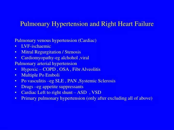
Pulmonary Hypertension and Right Heart Failure Pulmonary venous hypertension (Cardiac) • LVF-ischaemic • Mitral Regurgitation / Stenosis • Cardiomyopathy-eg alchohol ,viral Pulmonary arterial hypertension • Hypoxic – COPD , OSA , Fibr Alveolitis • Multiple Po Emboli • Po vasculitis –eg SLE , PAN ,Systemic Sclerosis • Drugs –eg appetite suppressants • Cardiac Left to right shunt – ASD , VSD • Primary pulmonary hypertension (only after excluding all of above)
Clinical Signs of Pulmonary Hypertension and Right Heart Failure • Central cyanosis if hypoxic • Dependent oedema • Raised JVP with V waves (due to secondary tricuspid regurg) • Right ventricular heave at left parasternal edge • Murmur of tricuspid regurgitation • Load P2 • Enlarged liver (pulsatile )
Investigation of Pulmonary Hypertension • ECG • CXR • SaO2 and arterial blood gases • Pulmonary function • Echocardiogram / Cardiac Catheterisation • D dimers and VQ scan if PE suspected • CT Pulmonary Angiogram • Auto-antibodies if vasculitis suspected
Primary pulmonary hypertension • Diagnosis by exclusion of other secondary causes • Progressive SOBOE and signs of right heart failure • Pharmacologic Treatment -prophylactic anticoagulation [warfarin] -O2 if hypoxic -Po Vasodilators :Endothelin antagonist (Oral Bosentan) , PDE5-inhibitor (Oral Sildenafil), iv Epoprostenol
THROMBOEMBOLIC LUNG DISEASE • Pulmonary infarction -in situ -venous emboli • Virchow’s Triad -Stasis -Vessel wall damage -Hypercoagulablity
RISK FACTORS FOR DVT AND PE #1 • Thrombophilia- FH,freq,site,age • Contraceptive pill ,HRT • Pregnancy • Pelvic obstruction-eg uterus,ovary,lymph nodes • Trauma-eg RTA
RISK FACTORS FOR DVT AND PE #2 • Surgery- eg pelvic,hip,knee • Immobility-eg bed rest,long haul flights • Malignancy • Myocardial infarction • Po hypertension/vasculitis
DVT • Proximal (Ileofemoral) -most likely to embolise -most likely to lead to chronic venous insufficiency and venous leg ulcers • Distal (Polpiteal) -least likely to embolise
Clinical presentation of DVT • Whole leg or calf involved depending on site • Swollen,hot,red,tender • Differential:Popliteal synovial rupture[Bakers cyst],Superficial thrombophlebitis,Calf cellulitis
Investigation of DVT • Ultrasound Doppler leg scan(1st line) -Non invasive -Exclude popliteal cyst, pelvic mass • CT scan of ileofemoral veins,IVC and pelvis • Constrast venography -Invasive,contrast(irritant,allergy) -Rarely indicated
Pulmonary Emboli • Predisposing DVT may be silent • Clinical presentation depends on size: • Large-cardiovascular shock,low BP,central cyanosis,sudden death • Medium-pleuritic pain,haemoptysis,breathless • Small recurrent-progressive dyspnoea, pulmonary hypertension and right heart failure
Diagnosis of PE #1 • Clinical Signs-Tachycardia,Tachypnoea,Cyanosis,Fever Low BP,Crackles, Rub, Pleural effusion • Arterial blood gases-PaO2,Sao2 (Type 1 resp failure:PaCO2 normal or low) • CXR-Normal early on before infarction -Basal atelectesis,Consolidation , Pleural effusion after infarction
Diagnosis of PE #2 Investigations • ECG :Acute Rt heart strain pattern (S1,Q3,T3 , T inv in V1-3) • D-dimers usually raised • Isotope lung scan (Ventilation/Perfusion) • Perfusion defect before infarction • Perfusion+Ventilation matched defect after infarction
V/Q isotope scan in Recurrent Po emboli Multiple filling defects (arrows) on perfusion (Q) scan Mismatched to ventilation (V) scan Dyspnoea ,Hypoxia,Cardiomegaly ,Po Hypertension and Large RV on Echo , Restrictive Lung Vols with Low DLCO ,Hypoxia
Diagnosis of PE #3 • CT pulmonary angiogram to image pulmonary artery filling defect • Leg and pelvic ultrasound to detect silent DVT • Echocardiogram to measure pulmonary artery pressure and RV size • Gas transfer factor (TLCO) to measure perfusion defect
CT Po Angiogram in Acute Massive PE Occluded Rt main Po Artery (arrow ) and filling defect Lt Po artery Acute Dyspnoea ,Hypoxia ,Low BP , Acute Rt Heart Strain on ECG Raised D dimers .No clot seen in IVC or ileofemoral veins Treated with Thrombolysis and Low MW Heparin
Investigation of underlying cause of PE • If no obvious underlying cause –eg surgery /pregnancy /malignancy /immobility • Look for underlying Ca – Clin exam ,CXR,PSA,CA125,CEA,Pelvic USS • Autoantibodies (SLE) – Antinuclear ,Anti-Cardiolipin • Coagulation factor screen – Antithrombin-3,Protein C/S, Factor 5/8
Prevention of DVT • Early post-op mobilisation • TED compression stockings • Calf muscle exercises • Subcutaneous low dose low mol wt heparin perioperatively • Dabigatran - direct thrombin inhibitor Rivaroxaban - direct inhibitor of activated factor X- both given orally for prophylaxis of venous thromboembolism in adults after hip or knee replacement surgery
Treatment of DVT/PE #1 • Anticoagulation prevents clot propagation-tips balance to thrombolysis-body dissolves clot • Initiate with parenteral heparin-fast acting-via antithrombin-3 • Usually therapeutic dose of s/c low mol wt heparin ( Dalteparin “Fragmin”)
Treatment of DVT/PE #2 • Low mol wt heparin –once daily injection ,no monitoring –no hassle • IV infusion unfractionated heparin -more hassle-need to monitor clotting, increased bleeding risk- rarely used nowadays
Treatment of DVT/PE #3 • Start concurrent oral warfarin-takes 3 days-antagonises vit K1 dependent prothrombin • After 3-5 days stop heparin-when INR>2 • Need to monitor APTT with unfractionated -but not with low mol wt heparin
Treatment of DVT/PE #4 • Continue Warfarin for 3-6 months • Monitor Warfarin with INR-Target range 2.5-3.5 • Interactions which increase anticoagulation -Alcohol,Antibiotics ,Aspirin,NSAIDs, Amiodarone, Cimetidine,Omeprazole ,etc etc • Look in BNF for possible interactions
Treatment of DVT/PE #5 • Thrombolysis-Streptokinase or TPA • Only for large life threatening PE-ie low BP and severe hypoxaemia due to main pulmonary artery occlusion • IVC filter to prevent embolisation from large ileofemoral/IVC clot - for recurrent PE’s • Thrombo-embolectomy –rarely indicated • Aspirin –no role – anti-platelet
Overanticoagulation • Address underlying cause-eg drug interaction,chronic liver disease,CHF • If bleeding then stop anticoagulant and reverse effect • Low MW Heparin has a long half life • Warfarin has a long half life • May need cover with prothrombin complex concentrate or fresh frozen plasma • Reverse warfarin with vitamin K1(especially if chronic liver disease) • Reverse heparin with protamine