Download
1 / 41
430 likes | 760 Views
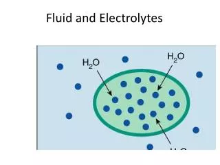
Fluid and Electrolytes. FLUIDS. Fluid homeostasis. Fluid homeostasis is dependent on Basal Inputs Vs. Outputs Our ability during pathologic processes to control Fluid loss Accounting for maintenance fluids Replacing ongoing loss Normal total body water

E N D
Fluid homeostasis • Fluid homeostasis is dependent on • Basal Inputs Vs. Outputs • Our ability during pathologic processes to control • Fluid loss • Accounting for maintenance fluids • Replacing ongoing loss • Normal total body water • It is also important to define what we mean by fluid? • Water Vs. Volume • Blood
Basal Input Vs. Output INPUT Oral intake ~ 2.5L Metabolic input ~ 200mls OUTPUT Urine 0.52ml/kg/hour IL insensible loss
Assessing fluid balance • Assessing fluid balance is SIMPLE • Be calculated and use all the tools available to you • General inspection • Physical examination • Adjuncts to the physical exam – daily weighs, inputs vs. outputs • Biochemistry
Assessing fluid balance • General inspection • Does the patient look well? • Sunken eyes • Kussmaul breathing • Conscious state • IV lines/catheter/NIBP/ CVP…. • Physical examination • Vital signs • BP + Postural • HR • RR • SaO2 • JVP – Efficacy? • Peripheral oedema • Pulmonary oedema • Cap refill
Assessing fluid balance • Adjuncts • Daily weighs • Assumes weight change is due to water change • Inputs Vs. Outputs • Oral/Parenteral Vs. Urine/Faeces/Vomitus/Drain outputs • Biochemistry • BUN:Creat ratio • Indicative of pre-renal dysfunction • Sodium • Can be used to assess total body water deficit/surplus • Based on normal TBW • Males 0.6% TB weight • Females 0.5% TB weight • Contraction alkalosis • ?Lactate?
In summary • General inspection • Physical examination • Adjuncts to the exam • Biochemistry
So after making you assessment what do you do? • FIRST Don’t meddle too much! Part of your assessment involves knowing when not to do anything • Is the process self-resolving/can it be treated by minimal input from yourself? • Is the patient DRY? Give them appropriate fluid • Is the patient WET? restrict input/remove fluid • Why are they dry/wet? How do we fix this process?
The DRY patient • What has made them dry as compared to baseline? • Reduced intake • Fluid loss • Two fluid rules of general medicine (geriatrics) • When the elderly become sick, they don’t drink • When they drink, they drink copious amounts of tea
Causes of fluid loss in the surgical patient • Fasting with inadequate replacement • Bowel prep • Pre/intra/post operative bleeding • Inappropriate post-operative hydration • Drains • Organ specific • GI – vomiting/diarrhoea/sequestration/NG aspirates/stoma loss • Urology – Post-obstructive diuresis • Neuro – Cerebral salt-wasting syndrome • Over-diuresis
Managing the fluid deficit patient; Fluid resuscitation • Replace losses bring the patient to euvolaemia • Based on weight • Fluid balance charts, estimated blood loss etc… • Biochemistry (will touch on this later) • Ensure adequate maintenance fluids ~ 2.5/3L/day • Replace ongoing losses • Look at drains • Urine output • ANY recordable ongoing output
Sometimes a fluid challenge is the best way to test if someone is DRY • Patients will not shatter, patients will not break • Be calculated and make a calculated decision
Fluid resuscitation in the shocked patient • Primary survey • Large bore IV canulas • 20ml/kg bolus • 500ml/1L bolus take into account left ventricular function • Re-assess fluid status post resuscitation to assess further need for resusc.
Estimating blood loss • Total blood volume • 66ml/Kg for males, 60mL/Kg for females • Class 1 <15% • Minimal signs due to transcapillary refill • Class 2 15%-30% • Orthostatic changes in HR/BP, some drop in U/O. Systemic vasoconstriction in play • Class 3 30% - 40% • Hypotension, low U/O, tachycardia, loss of systemic vasoconstriction • Class 4 >40% • U/O<5ml/hr, profound shock • When do I give blood? Acute phase vs. Delayed phase loss • Triggers? HCT? ScvO2?
FIX THE CAUSE • Large drain outputs • Massive haemorrhage • Etc… • Fluid will not always save the day • Never delay definitive treatment if it is indicated.
How do I give this patient fluid? • Oral • IV • Central line
What type of fluid do I use? • Depends on what you are trying to achieve • Increasing total body water? • 5% dextrose of 4% Dex and 1/5th N/S • Increasing total body volume? • Crystalloids/Colloids • Replacing blood loss • Blood • Augmenting cardiac ouput? • In the acidotic patient?
Just to clarify • Crystalloids • N/S, hartmann’s, 4% and 1/5th • Based on osmalarity 2/3rd will redistribute into the extravascular space so don’t get excited when your patient’s BP bumps up 5 mins after a fluid bolus! • Colloids • Gelofusine, albumin etc... • Theoretically to stay in the intravascular space…but for how long?
The WET patient • The WET patient • Too much input • Not enough output • OR the fluid is just in the wrong place • Distributive shock • Crystalloid resuscitation • Increased hydrostatic pressure with venous congestion • Decreased colloid oncotic pressure
Treating the wet patient • Do you need to? How much fluid O/L is tolerable? • Is this process unlikely to resolve? • Is the fluid load going to cause harm to the patient in the short term? • How to treat the wet patient • Fix the underlying cause • Fluid restriction • Diuresis • Physically removing the fluid paracentesis
So many electrolytes, so little time… • Hypernatraemia • Hyponatraemia • Hyperkalaemia • Hypokalaemia
Hypernatraemia • Can be due to Conn’s syndrome or excessive salt intake (sodium bicarbonate use etc…) • But… • Almost always water depletion • Treatment? • Calculate water deficit • cTBW x Current sodium = nTBW x normal sodium • nTBW = 0.6 x weight or 0.5 x weight • Replace water deficit • BD UEC’s to drop sodium by 0.5mmol/L/hour or ~10mmol sodium/day to prevent cerebral oedema
Hyponatraemia • Can occur in a • Hypovolaemic state • Euvolaemic state • Hypervolaemic state • Do a fluid assessment • Do the appropriate investigations
Evaluating hyponatraemia • Hypovolaemic • Urinary sodium • <20: Diarrhoea with Na loss and renal reabsorption • >20: Diuresis, Addison’s disease • Euvolaemic • Urinary osmolality (must compare to serum osmolality) • >100: SIADH • <100: Psychogenic polydipsia • Hypervolaemic • Urinary sodium • >20: Renal failure • <20: Heart failure
Correcting hyponatraemia • Fluid restriction<urine output • If Na change rapid, correct rapidly • Or correct rapidly within limits if patient symptomatic • If slow correct slowly • ?Sodium replacement? • Sodium deficit = nTBW x (130 – Current Plasma Na) • 3% sodium chloride contains 513 mEq of Na/L • Eg. 60kg woman with plasma Na 120 • Deficit = 0.5 x 60 x (130-120) = 300mEq • 300/513 = 585mL of hypertonic saline • Max rate correction of 0.5mmol/L/hr to prevent CPM
Hypokalaemia • Causes • Transcellular shift • Potassium depletion • Renal loss – diuretics, magnesium depletion (impaired K reabsorption) • Extrarenal loss – Diarrhoea • Treatment • 10% total body loss for each 1 mEq decrease • CHLORVESCENT • Slow K/IVT for longer term replacement (diuretics etc…) • Should we be scared of hypokalaemia? • It does not in itself produce serious ventricular cardiac arrhythmias but DOES increase arrhythmogenic potential of associated conditions (magnesium depletion, digitalis, myocardial ischaemia)
Biochemistry to diagnose hypokalaemia • Urine potassium • High >30mEq – renal loss • Cl >25mEq – diuretics, Mg depletion • Cl <15mEg – Ng drainage, alkalosis • Low <30mEq • Diarrhoea
Hyperkalaemia • Causes • Pseudohyperkalaemia – beware the 23g needle • Impaired excretion • Adrenal insufficiency, drugs (ACEi’s, spironolactone, NSAIDs) • Transcellular shift • Acidosis, rhabdo, drugs (B-blockers, digitalis) • Clinical manifestations • ECG changes ~ serum K of 6, definitely at 8
Hyperkalaemia - treatment • Treatment • Repeat bloods • ECG and monitoring • Membrane antagonism • Calcium gluconate Vs. Calcium chloride(x3) • Transcellularshift • Insulin/dextrose • B-agonists • Sodium Bicarbonate • Enhanced clearance • Resins • Diuretics • Haemodialysis