Download
1 / 55
570 likes | 711 Views
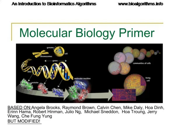
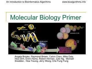
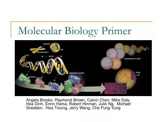
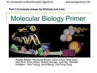
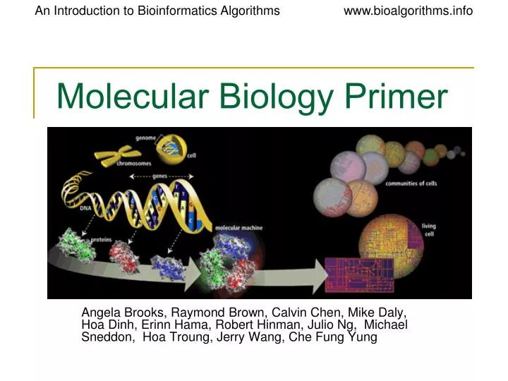
Molecular Biology Primer. Angela Brooks, Raymond Brown, Calvin Chen, Mike Daly, Hoa Dinh, Erinn Hama, Robert Hinman, Julio Ng, Michael Sneddon, Hoa Troung, Jerry Wang, Che Fung Yung. WHAT is a GENE?. Genes Make Proteins.

E N D
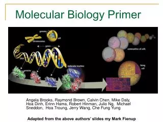
Molecular Biology Primer Angela Brooks, Raymond Brown, Calvin Chen, Mike Daly, Hoa Dinh, Erinn Hama, Robert Hinman, Julio Ng, Michael Sneddon, Hoa Troung, Jerry Wang, Che Fung Yung
Genes Make Proteins • genome-> genes ->protein(forms cellular structural & life functional)->pathways & physiology
Proteins: Workhorses of the Cell • 20 different amino acids • different chemical properties cause the protein chains to fold up into specific three-dimensional structures that define their particular functions in the cell. • Proteins do all essential work for the cell • build cellular structures • digest nutrients • execute metabolic functions • Mediate information flow within a cell and among cellular communities. • Proteins work together with other proteins or nucleic acids as "molecular machines" • structures that fit together and function in highly specific, lock-and-key ways.
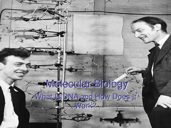
1 Biologist 1 Physics Ph.D. Student 900 words Nobel Prize Discovery of DNA • DNA Sequences • Chargaff and Vischer, 1949 • DNA consisting of A, T, G, C • Adenine, Guanine, Cytosine, Thymine • Chargaff Rule • Noticing #A#T and #G#C • A “strange but possibly meaningless” phenomenon. • Wow!! A Double Helix • Watson and Crick, Nature, April 25, 1953 • Rich, 1973 • Structural biologist at MIT. • DNA’s structure in atomic resolution. Crick Watson
Watson & Crick with DNA model Rosalind Franklin with X-ray image of DNA Watson & Crick – “…the secret of life” • Watson: a zoologist, Crick: a physicist • “In 1947 Crick knew no biology and practically no organic chemistry or crystallography..” – www.nobel.se • Applying Chagraff’s rules and the X-ray image from Rosalind Franklin, they constructed a “tinkertoy” model showing the double helix • Their 1953 Nature paper: “It has not escaped our notice that the specific pairing we have postulated immediately suggests a possible copying mechanism for the genetic material.”
DNA: The Basis of Life • Deoxyribonucleic Acid (DNA) • Double stranded with complementary strands A-T, C-G • DNA is a polymer • Sugar-Phosphate-Base • Bases held together by H bonding to the opposite strand
DNA, continued Sugar Phosphate Base (A,T, C or G) http://www.bio.miami.edu/dana/104/DNA2.jpg
DNA, continued • DNA has a double helix structure. However, it is not symmetric. It has a “forward” and “backward” direction. The ends are labeled 5’ and 3’ after the Carbon atoms in the sugar component. 5’ AATCGCAAT 3’ 3’ TTAGCGTTA 5’ DNA always reads 5’ to 3’ for transcription replication
DNA Components • Nitrogenous Base: N is important for hydrogen bonding between bases A – adenine with T – thymine (double H-bond) C – cytosine with G – guanine (triple H-bond) • Sugar: Ribose (5 carbon) Base covalently bonds with 1’ carbon Phosphate covalently bonds with 5’ carbon Normal ribose (OH on 2’ carbon) – RNA deoxyribose (H on 2’ carbon) – DNA dideoxyribose (H on 2’ & 3’ carbon) – used in DNA sequencing • Phosphate: negatively charged
Phosphate Sugar Basic Structure
Basic Structure Implications • DNA is (-) charged due to phosphate: gel electrophoresis, DNA sequencing (Sanger method) • H-bonds form between specific bases: hybridization – replication, transcription, translation DNA microarrays, hybridization blots, PCR C-G bound tighter than A-T due to triple H-bond • DNA-protein interactions (via major & minor grooves): transcriptional regulation • DNA polymerization: 5’ to 3’ – phosphodiester bond formed between 5’ phosphate and 3’ OH
The Purines The Pyrimidines
DNA - replication • DNA can replicate by splitting, and rebuilding each strand. • Note that the rebuilding of each strand uses slightly different mechanisms due to the 5’ 3’ asymmetry, but each daughter strand is an exact replica of the original strand. http://users.rcn.com/jkimball.ma.ultranet/BiologyPages/D/DNAReplication.html
Superstructure Lodish et al. Molecular Biology of the Cell (5th ed.). W.H. Freeman & Co., 2003.
Superstructure Implications • DNA in a living cell is in a highly compacted and structured state • Transcription factors and RNA polymerase need ACCESS to do their work • Transcription is dependent on the structural state – SEQUENCE alone does not tell the whole story
SWI/SNF SWI5 RNA Pol II TATA BP GENERAL TFs Transcriptional Regulation Lodish et al. Molecular Biology of the Cell (5th ed.). W.H. Freeman & Co., 2003.
The Histone Code • State of histone tails govern TF access to DNA • State is governed by amino acid sequence and modification (acetylation, phosphorylation, methylation) Lodish et al. Molecular Biology of the Cell (5th ed.). W.H. Freeman & Co., 2003.
Central Dogma of Biology The information for making proteins is stored in DNA. There is a process (transcription and translation) by which DNA is converted to protein. By understanding this process and how it is regulated we can make predictions and models of cells. Assembly Protein Sequence Analysis Sequence analysis Gene Finding
RNA • RNA is similar to DNA chemically. It is usually only a single strand. T(hyamine) is replaced by U(racil) • Some forms of RNA can form secondary structures by “pairing up” with itself. This can have change its properties dramatically. DNA and RNA can pair with each other. tRNA linear and 3D view: http://www.cgl.ucsf.edu/home/glasfeld/tutorial/trna/trna.gif
RNA, continued • Several types exist, classified by function • mRNA – this is what is usually being referred to when a Bioinformatician says “RNA”. This is used to carry a gene’s message out of the nucleus. • tRNA – transfers genetic information from mRNA to an amino acid sequence • rRNA – ribosomal RNA. Part of the ribosome which is involved in translation.
Terminology for Transcription • hnRNA (heterogeneous nuclear RNA): Eukaryotic mRNA primary transcipts whose introns have not yet been excised (pre-mRNA). • Phosphodiester Bond: Esterification linkage between a phosphate group and two alcohol groups. • Promoter: A special sequence of nucleotides indicating the starting point for RNA synthesis. • RNA (ribonucleotide): Nucleotides A,U,G, and C with ribose • RNA Polymerase II: Multisubunit enzyme that catalyzes the synthesis of an RNA molecule on a DNA template from nucleoside triphosphate precursors. • Terminator: Signal in DNA that halts transcription.
Transcription • The process of making RNA from DNA • Catalyzed by “transcriptase” enzyme • Needs a promoter region to begin transcription. • ~50 base pairs/second in bacteria, but multiple transcriptions can occur simultaneously http://ghs.gresham.k12.or.us/science/ps/sci/ibbio/chem/nucleic/chpt15/transcription.gif
DNA RNA: Transcription • DNA gets transcribed by a protein known as RNA-polymerase • This process builds a chain of bases that will become mRNA • RNA and DNA are similar, except that RNA is single stranded and thus less stable than DNA • Also, in RNA, the base uracil (U) is used instead of thymine (T), the DNA counterpart
Definition of a Gene • Regulatory regions: up to 50 kb upstream of +1 site • Exons: protein coding and untranslated regions (UTR) 1 to 178 exons per gene (mean 8.8) 8 bp to 17 kb per exon (mean 145 bp) • Introns: splice acceptor and donor sites, junk DNA average 1 kb – 50 kb per intron • Gene size: Largest – 2.4 Mb (Dystrophin). Mean – 27 kb.
Transcription: DNA hnRNA • Transcription occurs in the nucleus. • σ factor from RNA polymerase reads the promoter sequence and opens a small portion of the double helix exposing the DNA bases. • RNA polymerase II catalyzes the formation of phosphodiester bond that link nucleotides together to form a linear chain from 5’ to 3’ by unwinding the helix just ahead of the active site for polymerization of complementary base pairs. • The hydrolysis of high energy bonds of the substrates (nucleoside triphosphates ATP, CTP, GTP, and UTP) provides energy to drive the reaction. • During transcription, the DNA helix reforms as RNA forms. • When the terminator sequence is met, polymerase halts and releases both the DNA template and the RNA.
Central Dogma Revisited Splicing Transcription • Base Pairing Rule: A and T or U is held together by 2 hydrogen bonds and G and C is held together by 3 hydrogen bonds. • Note: Some mRNA stays as RNA (ie tRNA,rRNA). DNA hnRNA mRNA Nucleus Spliceosome Translation protein Ribosome in Cytoplasm
Terminology for Splicing • Exon: A portion of the gene that appears in both the primary and the mature mRNA transcripts. • Intron: A portion of the gene that is transcribed but excised prior to translation. • Lariat structure: The structure that an intron in mRNA takes during excision/splicing. • Spliceosome: A organelle that carries out the splicing reactions whereby the pre-mRNA is converted to a mature mRNA.
Splicing: hnRNA mRNA • Takes place on spliceosome that brings together a hnRNA, snRNPs, and a variety of pre-mRNA binding proteins. • 2 transesterification reactions: • 2’,5’ phosphodiester bond forms between an intron adenosine residue and the intron’s 5’-terminal phosphate group and a lariat structure is formed. • The free 3’-OH group of the 5’ exon displaces the 3’ end of the intron, forming a phosphodiester bond with the 5’ terminal phosphate of the 3’ exon to yield the spliced product. The lariat formed intron is the degraded.
Splicing and other RNA processing • In Eukaryotic cells, RNA is processed between transcription and translation. • This complicates the relationship between a DNA gene and the protein it codes for. • Sometimes alternate RNA processing can lead to an alternate protein as a result. This is true in the immune system.
Splicing (Eukaryotes) • Unprocessed RNA is composed of Introns and Extrons. Introns are removed before the rest is expressed and converted to protein. • Sometimes alternate splicings can create different valid proteins. • A typical Eukaryotic gene has 4-20 introns. Locating them by analytical means is not easy.
Terminology for Ribosome • Codon: The sequence of 3 nucleotides in DNA/RNA that encodes for a specific amino acid. • mRNA (messenger RNA): A ribonucleic acid whose sequence is complementary to that of a protein-coding gene in DNA. • Ribosome: The organelle that synthesizes polypeptides under the direction of mRNA • rRNA (ribosomal RNA):The RNA molecules that constitute the bulk of the ribosome and provides structural scaffolding for the ribosome and catalyzes peptide bond formation. • tRNA (transfer RNA): The small L-shaped RNAs that deliver specific amino acids to ribosomes according to the sequence of a bound mRNA.
Terminology for tRNA and proteins • Anticodon: The sequence of 3 nucleotides in tRNA that recognizes an mRNA codon through complementary base pairing. • C-terminal: The end of the protein with the free COOH. • N-terminal: The end of the protein with the free NH3.
Uncovering the code • Scientists conjectured that proteins came from DNA; but how did DNA code for proteins? • If one nucleotide codes for one amino acid, then there’d be 41 amino acids • However, there are 20 amino acids, so at least 3 bases codes for one amino acid, since 42 = 16 and 43 = 64 • This triplet of bases is called a “codon” • 64 different codons and only 20 amino acids means that the coding is degenerate: more than one codon sequence code for the same amino acid
RNA Protein: Translation • Ribosomes and transfer-RNAs (tRNA) run along the length of the newly synthesized mRNA, decoding one codon at a time to build a growing chain of amino acids (“peptide”) • The tRNAs have anti-codons, which complimentarily match the codons of mRNA to know what protein gets added next • But first, in eukaryotes, a phenomenon called splicing occurs • Introns are non-protein coding regions of the mRNA; exons are the coding regions • Introns are removed from the mRNA during splicing so that a functional, valid protein can form
Translation • The process of going from RNA to polypeptide. • Three base pairs of RNA (called a codon) correspond to one amino acid based on a fixed table. • Always starts with Methionine and ends with a stop codon
Translation, continued • Catalyzed by Ribosome • Using two different sites, the Ribosome continually binds tRNA, joins the amino acids together and moves to the next location along the mRNA • ~10 codons/second, but multiple translations can occur simultaneously http://wong.scripps.edu/PIX/ribosome.jpg
Protein Synthesis: Summary • There are twenty amino acids, each coded by three- base-sequences in DNA, called “codons” • This code is degenerate • The central dogma describes how proteins derive from DNA • DNA mRNA (splicing?) protein • The protein adopts a 3D structure specific to it’s amino acid arrangement and function
Proteins • Complex organic molecules made up of amino acid subunits • 20* different kinds of amino acids. Each has a 1 and 3 letter abbreviation. • http://www.indstate.edu/thcme/mwking/amino-acids.html for complete list of chemical structures and abbreviations. • Proteins are often enzymes that catalyze reactions. • Also called “poly-peptides” *Some other amino acids exist but not in humans.
Polypeptide v. Protein • A protein is a polypeptide, however to understand the function of a protein given only the polypeptide sequence is a very difficult problem. • Protein folding an open problem. The 3D structure depends on many variables. • Current approaches often work by looking at the structure of homologous (similar) proteins. • Improper folding of a protein is believed to be the cause of mad cow disease. http://www.sanger.ac.uk/Users/sgj/thesis/node2.html for more information on folding
Protein Folding • Proteins tend to fold into the lowest free energy conformation. • Proteins begin to fold while the peptide is still being translated. • Proteins bury most of its hydrophobic residues in an interior core to form an α helix. • Most proteins take the form of secondary structures α helices and β sheets. • Molecular chaperones, hsp60 and hsp 70, work with other proteins to help fold newly synthesized proteins. • Much of the protein modifications and folding occurs in the endoplasmic reticulum and mitochondria.
Protein Folding (cont’d) • The structure that a protein adopts is vital to it’s chemistry • Its structure determines which of its amino acids are exposed carry out the protein’s function • Its structure also determines what substrates it can react with
Major events in the history of Molecular Biology 1970 • 1970Howard Temin and David Baltimore independently isolate the first restriction enzyme • DNA can be cut into reproducible pieces with site-specific endonuclease called restriction enzymes; • the pieces can be linked to bacterial vectors and introduced into bacterial hosts. (gene cloning or recombinant DNA technology)