Download
1 / 60
600 likes | 701 Views
Visual System Examination. Dr Mahmood Fauzi Ansari MBBS MS FCLI Asst. Professor Ophthalmology Al Mareefa college of medicine 2013-14. Eyes are the window to the brain!. Before Visusal exam----WIPE. Wash hand Introduce self Patient name DOB

E N D
Visual System Examination Dr Mahmood Fauzi Ansari MBBS MS FCLI Asst. Professor Ophthalmology Al Mareefa college of medicine 2013-14
Before Visusal exam----WIPE • Wash hand • Introduce self • Patient name DOB • Explain examination and get consent
Learning objectives • Visual Acuity: CN II & beyond in the visual pathway • Visual Fields: Central & Peripheral Vision (retina to cortex) • Ocular Motility: (CN III, IV, VI) • Reflexes • Pupillary Light Reflex (CN II & III) • Accommodation (Near) Reflex • Fundus Examination (Retina including Optic disc):(Increased intracranial pressure; status of blood vessels reflecting hypertension, diabetes, etc.) • Visual Pathway: Since it is precisely organized & extends from the rostral to caudal aspect of the hemispheres, lesions along the pathway produce specific deficits that can aid in localizing the lesion.
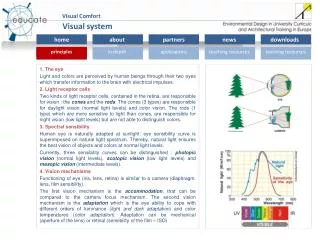
The Spectrum of Light 400-700um
The normal schematic eye Optic axis—The line passing through the centre of curvature of cornea and the two surfaces of the lens, meets the retina at fovea centralis. Nodal point—The optical centre lies in the posterior part of the lens. Anterior focal distance—It is about 15 mm in front of the cornea. Posterior focal distance—It is about 24 mm behind the cornea. The average power of a normal emmetropic eye is + 58 to + 60D. Most emmetropic eyes are approximately 24 mm in length.
What is Visual acuity? • The resolving power of the eye used to assess and quantify the eye’s ability to resolve varying letter sizes. • Visual acuity is a measurement of central vision only. • Assessment of total visual system from cornea to occipital cortex. • Visual acuity can be tested for both distance and near vision. • Distance visual acuity is the most common test. • Distance visual acuity (DVA) • 20ft or 6M is equivalent to optical infinity • Near visual acuity (NVA) • 40cm or 14 inches
Examples of visual acuity charts. (A) Snellen chart. (B) Landolt C chart. (C) Illiterate E chart.
Visual Acuity Grading (10th Revision of the WHO International Statistical Classification of Diseases). 1Good vision = 6/6 to 6/18. 2 Low vision = 6/24 to 3/60 (CF3m). 3Blind = 3/60 (CF3m) to PL (perception of light). 4Blind to light - NPL (no perception of light). • Legal blindness is defined as visual acuity (vision) of 20/200 (6/60) or less in the better eye with best correction possible. • In many areas, people with average acuity who nonetheless have a visual field of less than 10 degrees (the normal being 180 degrees) are also classified as being legally blind.
Near Visual Acuity • Testing the VA at close range (usually 40cm) • The purpose is to detect people with near vision difficulties (e.g., uncorrected high hyperopia, accommodative dysfunction) • In patients over 40 years old, the reduced near visual acuity is one of the symptoms of presbyopia Near Vision Charts Types -Reduced Snellen Acuity card Test distance at 14 inch (or 40cm) -Jaeger Acuity Card -Bailey-LovieReading Card -Point system -M notation
NEAR POINT • Near point is nearest possible distance at which the near object can be clearly seen. • It dramatically changes with age. • It is about 8 cm at age 10 and 100 cm at age 70 yrs 50 cm at the age of 50 yrs
Common causes of dimness of vision • Refractive error, cataract, glaucoma, corneal opacity, retinal pathology, optic atrophy
Refractive error • Refractive error is an optical abnormality of eye in which image is formed either infront or behind the retina. • Refraction is the procedure by which any refractive error is corrected with lenses. • Corrective lens is needed for proper focusing.
Types of refractive errors • EMMETROPIA -Normal optical condition of the eye. • AMETROPIA- ERRORS OF REFRACTION • Myopia (short-sightedness or near-sightedness) • Hyperopia (Long-sightedness or Far-sightedness) • Astigmatism • PresbyopiaInsufficiency of accommodation due to advancing age ( 40 years). • AphakiaAbsence of lens. It is a classical example of acquired high hypermetropia.
IMAGE FORMATION ON THE RETINA BEHIND THE RETINA INFRONT OR BEFORE RETINA
lenses Convex lens or plus lens Concave or minus lens Convex cylinder Concave cylinder
Image formation with/with out lens Hypermetropic eye Correction with convex lens
Color Sense • The normal colour vision is called “trichromatic” (red,green, blue) • COLOUR BLINDNESS [ACHROMATOPSIA] • an inability to recognisecolour. • Congenital—an inherited condition • Acquired—diseases of the macula and optic nerve, -Red color blindness (protanopia) -Green color blindness (deyteranopia) -Bluecolor blindness (tritanopia)
Pupil -should appear symmetric, -constrict to light (light reflex)
Afferent limb = Optic Nerve (SSA) Consensual Reflex Direct Reflex • Efferent limb = Oculomotor Nerve (GVE) • Postganglionic • Preganglionic Pupillary Constriction(Miosis) AKA Pupillary Light Reflex Right Left AFFERENT Retina Optic nerve Optic chiasmaOptic tract Pretectal Nucleus Parasympathetic Occulomotor nucleus EFFERENT Oculomotor Nerve Nerve to inferior obliqueCiliary ganglion Short ciliary nerve Sphincter pupillae and ciliaris muscle Nolte 17-38
Right Left B C Reflex abolished if afferent or efferent is damaged. Right Left Afferent defect Right Left Efferent defect Nolte 17-38
Cortex, Thalamus & Hippocampus Hypothalamus (CNS control center for ANS) ? ? Reticular Formation Reticulospinal fibers Preganglionic Sympathetic Neurons in Thoracic Cord (T1-T2) Superior Cervical Ganglion (post-ganglionic sympathetic) (pre-ganglionic sympathetic) Pupillary Dilation(Mydriasis) Decreased light to pupil Severe pain Strong emotional stimulus Dilation of pupil Horner’s Syndrome • Pupillary Constriction • Ptosis • Flushed & Dry Skin • Loss of Sympathetics • Lesion can be in CNS or PNS • Deficits ipsilateral to lesion
Ocular convergencePupillary constrictionLens thickening Accommodation (or “Near”) Reflex Shift in gaze from far to near. (contraction of pupil) 2. Three components: AFFERENT------ Retina Optic nerve Optic chiasmaOptic tract Lateral Geniculate Nucleus Optic Radiation Primary Visual Cortex EFFERENT----- -ParasympatheticOculomotorNuclei OculomotorNerve Ciliary ganglion Short ciliary nerve Sphincter pupillae and ciliaris muscle Marcus-Gunn pupil—There is ill-sustained contraction of the pupil in swinging flashlight test, e.g. as in retrobulbar neuritis. Argyll Robertson pupil: Pupillary constriction occurs as part of the accommodation reflex, but not in response to light.
PHYSIOLOGY OF VISION • In order to achieve clear vision, light reflected from objects within the visual field is focused on to the retina of both eyes. The processes involved in producing a clear image are: Refraction of the light rays • When light rays pass from a medium of one density to a medium of a different density they are refracted or bent. Accommodation of the eyes to light • 1. Pupil (constriction for near object and bright light • dilatation for distant object and dim light) • 2. Movement of the eyeballs-convergence • 3. Lens (flattened----far object / globular----near object)
FUNCTIONS OF THE RETINA • The retina is the photosensitive part of eye. The light sensitive cells are the rods and cones. • The rods are more sensitive than the cones. They are stimulated by low intensity or dim light, e.g. by the dim light in the interior of a darkened room (scotopic vision). • The cones are sensitive to bright light and colour. The different wavelengths of light stimulate photosensitive pigments in the cones, resulting in the perception of different colours. In a bright light the light rays are focused on the macula lutea(photopic vision). • Point of fixation—It is the area of maximum visual acuity in the normal visual field. It corresponds to the foveola of the retina. • Dark adaptation is the ability of the eye to adapt itself to decreasing illumination. Visual purple (rhodopsin) is a photosensitive pigment present only in the rods. The rate at which dark adaptation takes place is dependent upon the rate of reconstitution of rhodopsin.
1. Hemianopialoss of half the field of vision of both eyes 2. Amblyopiapartial loss of sight, fixation reflexes not developed. 3. Amaurosis complete loss of sight in one or both eyes LESIONS OF THE VISUAL PATHWAY
Inspection • Patient posture • Around bed walking aids, glasses • Eye inspections(Pupil size and symmetry Strabismus, Proptosis, Ptosis, Sclera, Around eye stye, swelling, discharge etc)
Monocular Visual Fields Temporal Field of Left Eye Lower Field of Left Eye Upper Field of Left Eye Nasal Field of Left Eye Horizontal Meridian Vertical Meridian UTQ UNQ F F LTQ LNQ Normal Monocular Visual Field of Left Eye Normal Monocular Visual Field of Right Eye Visual Fields Definition: The entire area that can be “seen” by the patient without movement of the head and with the eyes fixed on a single spot. • Mapping of Visual Fields: • Confrontational method • Perimetry (Manual or Automated) • Blind Spot • 15° to the temporal side of the visual field of each eye • On the horizontal meridian • Corresponds to the location of the optic nerve head 15° to the nasal side of the retina of each eye.
The Normal Field of Vision The optic chiasma
Perimetry • The term ‘perimetry’ is used to describe various techniques employed to evaluate both central and peripheral visual fields using targets of various sizes and colours. • Two techniques • Kinetic perimetry—A target is moved across the field to map out of the two-dimensional extent of field. • Static perimetry—Non-moving stimuli presented to obtain a vertical boundary / height of the visual field. Uses Charting of the visual fields is very useful in the diagnosis of many disease conditions • Glaucoma • Retinal diseases e.g. retinitis pigmentosa • Follow up of laser treatment for diabetic retinopathy • Neurological disorders, e.g. brain tumours, head injury, multiple sclerosis, cerebral thrombosis, aneurysms.
The Perimeter—(Lister’s, Goldmann’s) It consists of a half sphere within which a spot of light can be moved (kinetic technique). Method—The patient is seated with his chin supported by the chin rest. • One eye is covered by a pad. • The other eye fixes an object placed at the centre of the arc. • The field is recorded first with a white object 5 mm in diameter from periphery to centre. • At least 8 or preferably 16 meridians must be tested. Automated perimeters, e.g. Friedmannanalyser, Ouplot, Auto field perimeters Field master and Humphery field analyser Automated perimeters utilize computers to programme visual field sequences, e.g. Baylor visual field programmer attached to standard Goldmann perimeter.
Central Field (Campimetry) • Bjerrum’s screen—It consists of a black felt or flannel screen, 2 m in diameter on which • central 30° of the visual field can be studied (kinetic technique).
1.Electroretinogram (ERG) The changes induced by the stimulation of light in the resting potential of the eye are measured by electroretinography. It is extinguished or absent in complete failure of function of rods and cones, e.g. pigmentaryretinal dystrophy, complete occlusion of retinal artery, complete retinal detachment, advanced siderosis, etc. 2. Electro-oculogram (EOG) The changes in the resting current when the eyes are moved laterally are picked up by the electrodes placed at the inner and outer canthi. It is absent in retinal dystrophies and degenerations.
EXAMINATION OF THE FUNDUS OCULI • Pupil is dilated with a suitable mydriatic, e.g. phenylephrine, tropicamide, homide or cyclopentolateand the examination of the fundusoculi is done in a dark room. • Atropine is preferred in children as it results in paralysis of ciliary muscle.