Download
1 / 27
270 likes | 749 Views
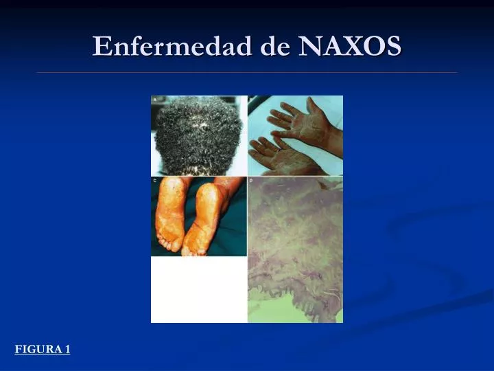
Enfermedad de NAXOS. FIGURA 1. MUTACIONES GENETICAS ASOCIADAS A MAVD. Tomé MT et al, Rev Esp Cardio 2004. FIGURA 2. Anatomia patologia. Corrado et al, Heart, 2000. FIGURA 3. Enfermedad EVOLUTIVA. 1.- Fase Temprana 2.- Fase Inestable 3.- Fase con fallo de VD. VI indemne

E N D
Enfermedad de NAXOS FIGURA 1
MUTACIONES GENETICAS ASOCIADAS A MAVD Tomé MT et al, Rev Esp Cardio 2004 FIGURA 2
Anatomia patologia Corrado et al, Heart, 2000 FIGURA 3
Enfermedad EVOLUTIVA 1.- Fase Temprana 2.- Fase Inestable 3.- Fase con fallo de VD. VI indemne 4.- Fase con fallo biventricular FIGURA 4
CRITERIOS DIAGNOSTICOS DE MAVD A pesarde todo se han encontradoafectacion exclusiva de VI. A pesarde todo se han encontradoafectacion exclusiva de VI. McKenna, et al. Diagnosis of ARVD/C. Task Force … Br Heart J 1994 FIGURA 5
Onda Epsilon FIGURA 6
Onda Epsilon FIGURA 7
Onda Epsilon FIGURA 8
Anchura de S ascendente > 55 ms FIGURA 9 Nasir et al, Electrocardiographic Features of ARVD/C According to Disease Severity. Circulation 2004
TVMS FIGURA 10
TVMS FIGURA 11
Ecocardiograma FIGURA 12
Ventriculografía FIGURA 13
Aneurismas en VD FIGURA 14
Dilatación del TS de VD FIGURA 15
ESTRATIFICACION DEL RIESGO Necropsia MAVD Evolución FV Evolución FIGURA 16
Mortalidad por enfermedades del miocardio en niños y jóvenesResultados: Datos clínicos FIGURA 17
ESTUDIOS NECROPSICOS FIGURA 18
Hulot et al, Natural History and Risk Stratification of ARVD/C. Circulation 2004 FIGURA 19
Hulot et al. Circulation 2004 FIGURA 20
Corrado et al, ICD Therapy for Prevention of SD in Patients With ARVC/D. Circulation 2003 FIGURA 21
MARCADORES DE FV • Parada Cardiorespiratoria (P 0.007) • TV con afectación hemodinamica (P 0.037) • FE disminuida en VI (P<0.0001) • Descubrimiento de la enfermedad en edad temprana (P 0.015) • Síncope inexplicado (P 0.07) Corrado et al, Circulation 2003 FIGURA 22
TRATAMIENTO • Farmacologico • Ablación con Radiofrecuencia • Implante de un Desfibrilador Antitaquicardia • Trasplante FIGURA 23
DIAGNOSTICO DIFERENCIAL TVTSVD versus MAVD FIGURA 24 Della Bella, Heart 2000
ABLACION CON RADIOFRECUENCIA FIGURA 25
Marchlinski, Circulation 2004 FIGURA 26
Corrado et al, Circulation 2003 FIGURA 27