Download
1 / 52
520 likes | 842 Views
Hemangiomas and Vascular Malformations. Yağmur AYDIN, M.D. University of Istanbul , Cerrahpasa Medical Faculty Department of Plastic , Recons t. and Aesthetic Surgery. Vascular Anomalies. In U.S, 40000 babies with vascular anomailes are born in a year

E N D
HemangiomasandVascularMalformations Yağmur AYDIN, M.D. University of Istanbul, CerrahpasaMedicalFacultyDepartment of Plastic, Reconst. and Aesthetic Surgery
Vascular Anomalies • In U.S, 40000 babieswithvascularanomailesareborn in a year • 1of 10 childrenhas vascularanomaly • A commonmistakehas been done withthenaming • Strawbery, cavernous, capillaryhemangioma • MullikenandGlowacki in 1982 – classificationaccordingtothebiologicalcharacteristics(endothelialproperties)
Vascular Anomalies • Tumors • Hemangioma • pyogenicgranuloma • Kaposiphormhemangio-endothelioma • Malformations • Capillary • Lymphatic • Venous • Arteriovenous • combined
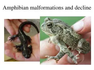
Hemangioma • Themostcommontumor of infancyandchildhood (4-10%) • 3-5 timesmoreseen in girls • Moreseen in prematureinfants (<1200 grams% 23) • Not frequent in darker-skinnedbabies • Usuallyoccurs in first 2 weeksafterbirth • Initially, a pale-colored, telangiectaticormacularredstainorpurple-coloredstain • Singlelesion in 80%, 20% morethanonelesion • Inpatientswithmorethanonelesionaccompaniesothersystemhemangiomas( liveretc.)
ClinicalAppearance • Lesions located in the superficial dermis • Hard, shiny dark-red coloured bulky lesion • Lesion located in deep dermis, subcutaneous fat or muscle • Showing a slight bulking, hot, bluish coloured lesion
The incidence of hemangiomas • Craniofacial area (60%) • The body (25%) • Extremities (15%)
3 phases of hemangiomas • Proliferationperiod (0-1 years) • Rapidlydividingendothelialcells • İnvolutionperiod (1-5 years) • Reducedendothelialproliferation, increasedapoptosis, fibrofattyreplacement, reducedtumorvolume, skinsoftening • Involutioncompletionterm (> 5 years) • Thinveinsandcapillariesthatdraincurrentfibrofatty islets
Proliferation period: • Rapid growth (upto10-12. months) • Involution period (1-7 years) • Growth slows down,compatiblewith the child's growth rate • Fading of the skin starting from the center of the lesion,softening • The color disappearsuntill 5-7 years • Normal skin takesplace in nearly50% of children
Differential Diagnosis • Lymphatic malformation –deep located hemangioma (neck, axilla) • Capillary malformation - macular hemangioma • Vascular tumors of infancy - Fibrosarcoma etc. • Radiological studies • Biopsy
Clinical evaluation • Clearand open dialoguewith the family • Confirmthe diagnosis • Documentation with photos • The need for medicalorsurgicaltreatment • Other diagnostic studies to investigate the extensionofhemangiomas and otheranomalies • Provide support groups andpublications forfamily
Complications • Ulceration, bleeding • Infection • Visualimpairment • Airwayobstruction • Obstructionin theearcanal • Congestiveheartfailure • diffuseneonatalhemangiomathosis • largevisceralhemangiomas
Problems • Hemangiomaslocated in head&neck(PHACES syndrome.) • Hemangiomascoveringthevisualarea • Periorallocation • Respiratory distress (subglottichemangioma • Ulcer
hemangiomacoveringthevisualfield in 2 monthsoldinfant Rapidreductionafter oral andintralesionalcorticosteroidinjection Residuemass at age 1,atrophy, telangiectasia
Treatment • Followupandobservation • Trainingandconvincingthefamily • Systemiccorticosteroid • Second-generationdrugs (vincristine, interferon alpha) • Lasertherapy - pulsed-dyelaser • ulceratedhemanjyom • Remainingtelangiectasiaafterinvolution
Hemangiomas requiring treatment • Coveringthe visualarea, the air way, the ear canal • Leading to congestive heart failure • Showing ulceration and bleeding
Treatment • Dangerous and life-threatening complicationsoccur in 10% of patient • The first option in medical treatmentapplication of corticosteroids (90% response) • Corticosteroid • Topical / injection into the lesion • Triamcinolone 3-5 mg / kg • 3-5 timesapplication (6-8 weeks apart) • systemic application • Oral prednisone or prednisolone 2-3 mg / kg /day • Once every 2-4 weeks (10-12 months)
Othermedicaltreatmentagents(vicristine, interpheronalpha) • Casesnon –responsiveto corticosteroid therapy • If long-term use of corticosteroids is contraindicated • Ifcomplicationsdevelopafterthe use of corticosteroids • Incasesthatthe family does not want to use (rare)
Ulcer local diffuse
Ulcer Treatment • Dressings • Corticosteroid • Laser( flashlamp dye laser) • Total excision (if primary closure is possible)
residual athrophic • tissue and wrinkled skin after involution • of hemangioma
Clinical appearance afterinvolution of a largeperioralandperiorbitalhemangioma
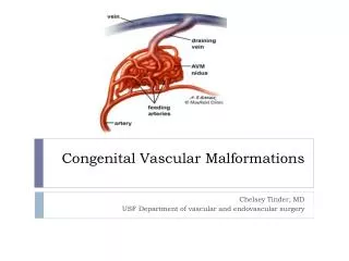
Vascular Malformations • As a result of an error during embryological andfetal development • Classification • Clinical • Radiological • Histological
Vascular Malformations • Capillary • Lymphatic • Venous • Combined • Arteriovenous • Lymphatico-venous • Lympathico-capillary-venous
Vascular Malformations • Slow-Flow • Capillary • Lymphatic • venous • Fast-flow • Arteriovenous
Capillary Malformations • "Port wine stain“ • present at birth • Pink or red-colored intradermal discoloration • Small, or large enough to cover the extremity or the face • Seen in 3 infants per 1000 live births
Real-capillary malformations • Progressive • Growthicker over time, darken and noduledevelopsinside • "Salmon patch", "Nevus simplex", "vascularstain ” “birthstain” • Oftenseen in the middle part of the face, neck • Generallyfade andreduce at theage of 1
Otherunderlyingdiseaseorsyndrome • Capillarymalformation on midlinelumbarorcervicalregion,(spinaldysraphism, tetheredspinalcord) • Sturge-Webersyndrome (capillarymalformation of thedistributionarea of thetrigeminalnerve,lepthomeningealvascularabnormalities,seizures) • Klippel-Trenaunaysyndrome(slow- flowcapillary-lympho-venousmalformation , elongation on axialplaneandovergrowth of a limb
Sturge-Webersyndrome • capillary malformation of the trigeminal nervedistribution area • lepthomeningeal vascular anomaly • Seisures
Klippel-Trenaunay syndrome • capillary malformation • soft tissue and bone overgrowth • varices
Treatment • Flashlamp-pumpedpulseddyelaser • 577, 585, or 595 nmwavelength • Targetsoxyhemoglobin • Provokesintravascularthrombosis • 50- 90% of patientspresentdiscoloration • Treatmentgivesthebestresults in earlychildhood • Otherlasers (non-responsivepatients) • Alexandrite (755 nm) • Neodymium: yttrium-aluminum-garnet (1064nm) • Intensepulsedlight (IPL)
Port wine stainThe result obtained after two applications of pulsed dye laser
Lympathic Malformations • Seen as local sponge-like lesionsordifuse lesions covering an anatomical organ orarea • Radiological and histological • Microcystic • Macrocyctic • Mixed • Usually occurs at birth or first 2 ages • most common seen in cervicofacialarea • Axilla, chest, mediastinum, retroperitoneal area,perineum, glutealarea • Overlying skin is intact, looksbluish • Small ,thin vesicles arepathognomonicfordermal involvement
Treatment • Bleeding • Recurrentinfection (cellulitis) • Body contouring • Correction of funtionaldeficits • Sclerotherapy (macrocycticlesions) • Intralesionalinjection of bleomycin • OK-432 • Argon, neodynium: YAG, orcarbondioxidelaser
Surgical treatment indications • Lesions blocking the respiratoryway • Lesions that create feeding problems • Lesions making distortion
Venous Malformation • Soft, fadingwith pressure, bluish coloured masses under the skin • Swellingwith physical activity and whenslouched down • Palpable thrombi • Morningpain (stasis and microthrombi) • Frequent localization of head and neck • Moreexpansive than itlooks(muscle,bone, oral mucosa, salivary gland)
Diagnosis • Magnetic resonance imaging (MRI) • To diagnose • To evaluate the extent of malformation • Bleeding-coagulation profile should be assessed • coagulopathy
Treatment • Percutaneoussclerotherapy • absoluteethanol • hypertonicsaline • sodiumsulfatetetradesil • Elasticcompressionstockings (extremity) • Aspirin (daily) • painfulthrombus • Forprophylaxis of phlebitis • SurgicalTreatment • headandnecklesionscausingcosmetic problem • severe painandbleeding • Lesionswithwell-definedborders
Venous malformation spreading into the vulva and inner thigh muscles Partial excision and injection of sclerosing agents
Venous malformation: 2 months after1 time the Nd: YAG laser application • complete regression
Arterio-venous malformations • Connection exists between the arterial system and venous system • They usually present at birth • Often incorrectly diagnosed (KM orhemangioma) • There is a rapidflow between the two systems • Fast flow is evident in childhood
Arterio-venous malformations • Over time the stain on the skin • Erythema • The local rise of temperature • Thrill • Murmur • The mass may expand • Rapid growth may be seen during puberty or trauma
Arterio-venous malformations • Arteriovenous shunts • Ischemia and related symptoms and signs • painless ulceration • Persistent pain, occasional bleeding • Widespread AVM may increasecardiac output and cause congestive heart
Diagnosis • Ultrasonography • ColouredDoppler • MRI • Angiography • Feederanddrainingvessels • Varyingdegrees of arterialdilatation • curvedvessels • A-V shunts • Enlargeddrainingveins
Treatment • Embolization • Sclerotherapy • Surgical resection and reconstruction • Preoperative angiography • Determines feeder and draining vessels • Embolization (adhesive or with special springs)
Arterio-venous malformations • Intracranial (most common) • Extracranial • Head and neck • Extremity • Body • İnternal organs