Download
1 / 32
340 likes | 859 Views
Cholesteatoma-Pathogenesis and Surgical Management. Grand Rounds Presentation February 24, 1999 Kyle Kennedy, M.D. Jeffrey Vrabec, M.D. Introduction. Cholesteatoma (keratoma)-essentially an accumulation of skin in ME/mastoid insidious nature

E N D
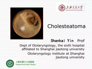
Cholesteatoma-Pathogenesis and Surgical Management Grand Rounds Presentation February 24, 1999 Kyle Kennedy, M.D. Jeffrey Vrabec, M.D.
Introduction • Cholesteatoma (keratoma)-essentially an accumulation of skin in ME/mastoid • insidious nature • variable symptoms depending on extent and location of disease • primarily a surgical disease • high rate of recidivistic disease • long-term follow-up essential
Introduction • Pathology and classification • Eustachian tube dysfunction • Pathogenesis • Anatomic considerations • Evaluation • Surgical management • Results of therapy • Complications
Pathology and Classification • Non-neoplastic accumulation of keratinizing stratified squamous epithelium with desquamated keratin debris • Subepithelial fibroconnective tissue • Granulation tissue • Bone destruction possible • Elaboration of collagenase and other inflammatory mediators
Pathology and Classification • Congenital cholesteatoma • Acquired cholesteatoma • Canal cholesteatoma
Congenital Cholesteatoma • Cholesteatoma sac medial to an intact tympanic membrane • Normal pars flaccida and tensa • No h/o TM perforation or otorrhea • No h/o otologic trauma or surgery • H/o prior episodes of OM does not preclude its presence
Acquired Cholesteatoma • Usually found in posterosuperior quadrant of TM with asso. retraction pocket or perforation • Primary acquired cholesteatoma asso. with pre-existing retraction pocket • Secondary acquired cholesteatoma arises in setting of persistent TM perforation
Canal Cholesteatoma • Found lateral to TM • Idiopathic, post-traumatic, and iatrogenic variants • Must be distinguished from keratosis obturans
Eustachian Tube Dysfunction • Important in pathogenesis of middle ear disease and cholesteatoma • Essential role in recurrent disease and surgical failure • Preoperative clinical assessment of tubal patency mandatory • Tubal function and ME aeration particularly important in postoperative hearing results
Pathogenesis • Migratory nature of TM epithelium and cholesteatoma • Iatrogenic implantation • Invasion of squamous epithelium • Invagination theory • Basal cell proliferation • Metaplasia • Embryonic squamous epithelial cell rests
Anatomic Considerations • Tympanic cavity derived from endodermally-lined first branchial pouch • Characteristic pathways of disease spread • Attic or epitympanum-Prussack’s space • Posterior mesotympanum-facial recess and sinus tympani
Evaluation • History-long h/o ear complaints • Physical examination-otomicroscopy • Audiology-CHL • Imaging-assessment of mastoid disease, surgical road map, revision cases, sensorineural hearing loss, vestibular symptoms
Management • Surgical disease • Patient age (I.e. pediatric cholesteatoma generally considered more aggressive) • Primary goal is eradication of disease with hearing preservation or improvement secondary • Final therapeutic decisions often made at surgery
Non-surgical Management • Office management of limited disease in elderly patients with comorbidities • Topical antibiotic preparations including those containing steroids sometimes useful preoperatively
Surgical Management • No consensus regarding optimal surgical strategy • Principal controversy concerning intact canal wall vs. canal wall down mastoidectomy • Therapy must be individualized on case-by-case basis
Preoperative Patient Counseling • Surgical goals • Risks of surgery including facial paralysis, tinnitus, vertigo, worsening of hearing • Possible need for staged procedure • Chronic nature of disease process with need for long-term follow-up • Routine aural toilet if mastoid bowl created
Tympanostomy Tube Insertion • Alleviation of early TM retraction in setting of ETD • Arrest pathologic process prior to irreversible changes such as atelectasis, deep retraction pocket formation, TM perforation, or cholesteatoma formation • Assist in maintenance of ME aeration after tympanoplasty or tympanomastoidectomy
Tympanomeatal Flap/Tympanoplasty • Smaller congenital cholesteatomas of involving TM or ME • Acquired cholesteatomas limited to mesotympanum
Intact Canal Wall Mastoidectomy • Preservation of posterior canal wall during simple mastoidectomy with or without posterior tympanotomy (facial recess approach) • Cholesteatomas of attic, antrum, post. mesotympanum with adequate ME and mastoid aeration • Staging necessary with ME mucosal abnormalities, ossicular erosion, residual disease
Canal Wall Down Mastoidectomy • Removal of post. canal wall to level of vertical facial nerve • Creation of mastoid cavity with exteriorization of mastoid into EAC • Scutum removed with obliteration of epitympanum and removal of malleus head and incus • MRM ME space maintained while radical mastoid eliminates ME space and obliterates eustachian tube
Canal Wall Down Mastoidectomy • Surgery in an only-hearing ear • Poor anesthetic risk • Poor pt compliance with unreliable F/U • Poor tubal function and ME aeration • Sclerotic mastoid • Extensive canal wall defect • Labyrinthine fistula • Meatoplasty and mastoid obliteration
Atticotomy • Removal of scutum • Limited attic disease • Scutal reconstruction with autologous cartilage
Bondy Procedure • Removal of scutum and posterior canal wall with preservation of ossicles and ME space • Larger attic cholesteatomas lateral to ossicles in pt with sclerotic mastoid
Intact Canal Wall Advantages • More rapid healing • Easier long-term postoperative care • No water precautions necessary (particularly important in children) • More options available for hearing aid, if necessary
Intact Canal Wall Disadvantages • Epitympanum/mastoid not accessible to postop inspection • Supratubal space not easily accessible unless malleus head and incus removed • Both residual and recurrent disease more likely • Greater number of procedures usually required for disease eradication
Canal Wall Down Advantages • Easy detection of residual disease • Recurrent cholesteatoma rare • Fewer procedures necessary for eradication of disease
Canal Wall Down Disadvantages • Longer healing time • Special cavity care often necessary for proper healing • Periodic cleaning necessary • Accumulation of debris may occur with increased risk of infection • Water precautions necessary
Results of Therapy • Rosenberg et al. examined variables with regard to residual-recurrent disease (retrospective) • 232 children with cholesteatoma (244 ears) • Ossicular erosion asso. with residual-recurrent disease (necessitates 2nd look) • Recidivism 61% at 6 years (necessitates long-term F/U)
Results of Therapy • Dodson et al. examined cases of 66 children with cholesteatoma (73 ears) retrospectively with ave. F/U 37.7 mos. • ICW-41% recidivism and CWD-12% recidivism • Postop SRT less than 30 dB in 75% of ICW and 72% of CWD • Prefer ICW with 2nd stage
Results of Therapy • Hirsch et al. retro. reviewed 164 cases of ped. chol. (116 avail. for 5 year F/U) • Majority of pts required CWD procedure • Recidivism 11% for tympanoplasty, 19% for ICW, 5% for MRM, and 0% for radical mastoid • Also reported fewer revisions and better hearing results with CWD
Complications • Conductive hearing loss • Labyrinthine fistula • Facial nerve paresis or paralysis • Intratemporal or intracranial complications • Encephalocele
Conclusions • Exact pathogenesis not entirely clear • Important anatomic considerations in management • Eradication of disease primary goal • No universally accepted surgical strategy • High rate of recidivism with long-term F/U essential • Maintain vigilance for complications