Download
1 / 17
240 likes | 964 Views
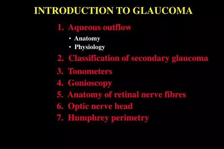
INTRODUCTION TO GLAUCOMA. 1. Aqueous outflow. Anatomy. Physiology. 2. Classification of secondary glaucoma. 3. Tonometers. 4. Gonioscopy. 5. Anatomy of retinal nerve fibres. 6. Optic nerve head. 7. Humphrey perimetry. Aqueous outflow. Anatomy. Physiology. a - Uveal meshwork.

E N D
INTRODUCTION TO GLAUCOMA 1. Aqueous outflow • Anatomy • Physiology 2. Classification of secondary glaucoma 3. Tonometers 4. Gonioscopy 5. Anatomy of retinal nerve fibres 6. Optic nerve head 7. Humphrey perimetry
Aqueous outflow Anatomy Physiology a - Uveal meshwork a - Conventional outflow b - Corneoscleral meshwork b - Uveoscleral outflow c - Schwalbe line c - Iris outflow d - Schlemm canal e - Collector channels f - Longitudinal muscle of ciliary body g - Scleral spur
Classification of secondary glaucomas Open-angle a b a. Pre-trabecular - membrane over trabeculum b. Trabecular - ‘clogging up’ of trabeculum Angle-closure c d c. With pupil block - seclusio pupillae and iris bombé d. Without pupil block - peripheral anterior synechiae
Tonometers Goldmann Schiotz Perkins Contact applanation Contact indentation Portable contact applanation Pulsair 2000 (Keeler) Tono-Pen Air-puff Portable non-contact applanation portable contact applanation Non-contact indentation
Goniolenses Goldmann Zeiss • Single or triple mirror • Four mirror • Contact surface diameter 12 mm • Contact surface diameter 9 mm • Coupling substance required • Coupling substance not required • Suitable for ALT • Not suitable for ALT • Not suitable for indentation gonioscopy • Suitable for indentation gonioscopy
Indentation gonioscopy Differentiates ‘appositional’ from ‘synechial’ angle closure Press Zeiss lens posteriorly against cornea Aqueous is forced into periphery of anterior chamber
Indentation gonioscopy in iridocorneal contact During indentation Before indentation • Part of angle is forced open • Complete angle closure • Apex of corneal wedge not visible • Part of angle remains closed by PAS
Angle structures Schwalbe line Trabeculum Schlemm canal Scleral spur Iris processes
Shaffer grading of angle width Grade 4 (35-45 ) • Ciliary body easily visible Grade 3 (25-35 ) • At least scleral spur visible Grade 2 (20 ) 2 3 1 • Only trabeculum visible 4 0 • Angle closure possible but unlikely Grade 1 (10 ) • Only Schwalbe line and perhaps • top of trabeculum visible • High risk of angle closure Grade 0 (0 ) • Iridocorneal contact present • Apex of corneal wedge not visible • Use indentation gonioscopy
Anatomy of retinal nerve fibres Papillomacular bundle Horizontal raphe
Optic nerve head Small physiological cup a - Nerve fibre layer a b b - Prelaminar layer c - Laminar layer c Large physiological cup • Normal vertical cup-disc ratio is 0.3 or less • 2% of population have cup-disc ratio > 0.7 • Asymmetry of 0.2 or more is suspicious Total glaucomatous cupping
Types of physiological excavation Cup with sloping temporal wall Larger and deeper punched-out central cup Small dimple central cup
Pallor and cupping Pallor - maximal area of colour contrast Cupping - bending of small blood vessels crossing disc Cupping and pallor correspond Cupping is greater than pallor
Reliability Indices 1. Fixation losses • Detected by presenting stimuli in blind spot 2. False positives • Stimulus accompanied by a sound • High score suggests a ‘trigger happy’ patient 3. False negatives • Failure to respond to a stimulus 9 dB brighter than previously seen at • same location • High score indicates inattention, or advanced field loss
Deviations 1. Total • Upper numerical display shows difference (dB) between • patient’s results and age-matched normals • Lower graphic display shows these differences as grey scale 2. Pattern • Similar to total deviation • Adjusted for any generalized depression in overall field
Global Indices 1. Mean deviation (elevation or depression) • Deviation of patient’s overall field from normal • p values are < 5%, < 2%, < 1% and < 0.5% • The lower the p value the greater the significance 2. Pattern standard deviation • Departure of visual field from age-matched normals 3. Short-term fluctuation • Consistency of responses • 2 dB or less indicates reliable field • > 3 dB indicates either unreliable or damaged field 4. Corrected pattern standard deviation • Departure of overall shape of patient’s hill of vision from • age-matched normals