Download
1 / 26
260 likes | 281 Views
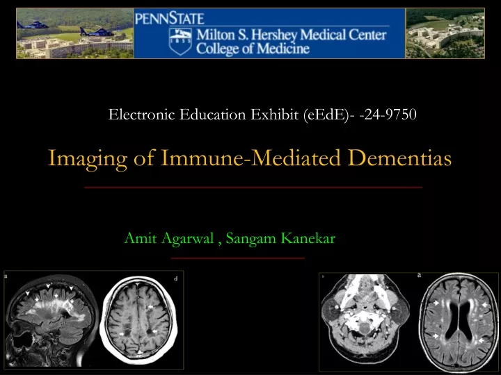
Electronic Education Exhibit ( eEdE )- - 24-9750 Imaging of Immune-Mediated Dementias. Amit Agarwal , Sangam Kanekar. Statement of Disclosure.

E N D
Electronic Education Exhibit (eEdE)- -24-9750 Imaging of Immune-Mediated Dementias Amit Agarwal , Sangam Kanekar
Statement of Disclosure Neither the author nor co-authors have any financial interest or other relationship with the manufacturer(s) of any commercial product(s) or services discussed in this electronic presentation
Contents • Introduction • Categories of Immune-mediated Dementias • Diseases with specific antigen/antibodyParaneoplastic neurologic disorders Multiple sclerosis Hashimoto's encephalopathy (HE) Gluten sensitivity (GS) dementia Systemic lupus erythematosus (SLE) Sjögren'sencephalopathy dementias Autoimmune-mediated channelopathies Anti–glutamic acid decarboxylase [anti-GAD] syndrome • Diseases without specific antigen/antibody Behçet's disease Sarcoidosis Primary angiitis of the central nervous system (PACNS) • Conclusion
Introduction • A myriad of autoimmune disorders can affect brain, and it would be impossible to encompass them in a single review. • We will focus on the most prevalent immune mediated cognitive decline and dementia. • The hallmarks of these disorders are: • -Rapidly progressive fluctuating course • -Detection of autoantibodies in the peripheral blood or CSF • -Indications of inflammation in the CSF such as pleocytosis, elevated protein level, and increased immunoglobulin G index * Teaching Point: Myriad of autoimmune disorders can affect brain and present with dementia, usually with a rapidly progressive course. Auto-antibodies are detected in CSf
Role of Imaging • The traditional view has been that computed tomography (CT) and magnetic resonance imaging (MRI) are performed to exclude other abnormalities that are potentially amenable to surgical treatment, such as a tumor, hematoma, or hydrocephalus • However, in the recent practice parameter on the diagnosis of dementia, structural neuroimaging in the routine initial evaluation of patients with dementia is recommended as a guideline. • Functional imaging provides insight into the operational aspects of the brain, and since it appears that brain pathology in dementia begins long before there is clinical evidence of disease, functional imaging is attractive for the early detection of dementia. Single-photon emission computed tomography (SPECT), positron emission tomography (PET), and functional MRI (fMRI) are becoming increasingly relevant to the study of dementia
Multiple Sclerosis • MS remains a clinical diagnosis and is supplemented by imaging and CSF findings with axonal and neuronal degeneration in the early stages of the disease. • Besides the sensory and motor dysfunction exhibited in MS, cognitive impairment occurs in 40–70% of patients. Cognitive impairment can occur early in the course of MS and is sometimes the first manifestation of the disease. • The cortex, the subcortical region, and the fiber’s integrity in the white matter are essential for the brain’s cognitive functioning. • Cortico-subcortical connections, controlling the information processing speed, and interlobarconnections, responsible for attention, memory and executive processes may be affected in MS by various cortico-subcortical, perivascular, intracortical and /or band-like subpial demyelinating lesions. • Specific domains of cognitive functioning appear particularly susceptible in MS. Learning, memory, conceptual reasoning, speed of information processing, attention, and executive functioning are most frequently affected. Language, semantic memory, and attention span are rarely involved
Multiple Sclerosis • Traditional T2-weighted MRI techniques may not reflect the full extent of neuronal tissue damage in MS. Nonenhancing black holes on T1 weighted images correlates well with axonal damage in histopathology [Figure1] • These white matter lesions cause disruption of neural connections among cortical associative areas as well as between cortical and subcortical structures. This forms the basis of the MS dementia • Whole brain atrophy is one of the strong markers for cognitive decline in MS [Figure2]. Cortical gray matter, central atrophy (particularly the atrophy of the thalamus) and dilatation of the third ventricle are correlated with cognitive impairment in MS • N-acetyl aspartate (NAA), a marker for the neuronal and axonal integrity is decreased in all states and types of MS with corresponding increase in the choline, myoinositol, and creatine indicating glial proliferatio * Teaching Point: Traditional T2-weighted MRI techniques may not reflect the full extent of neuronal tissue damage in MS. Whole brain atrophy is one of the strong markers for cognitive decline in MS
Multiple Sclerosis AJR 2010; 195:W164–W171 Radiographics 2000: 20:83-98 Fig1. (A) Sagittal FLAIR and axial T1W images show extensive focal and diffuse lesions in the cerebral white matter with predilection for the periventricular region (arrows in sagittal image). Many of these lesions are hypointense on T1WI indicating tissue destruction and are called as “black holes” (arrows in T1 axial). Also note generalized atrophy of the cerebral cortex (arrowheads).
Multiple Sclerosis AJR 2010; 195:W164–W171 Radiographics 2000: 20:83-98 Fig2. (A) Axial and (B) sagittal FLAIR images of MS patient (from 2004 MRI) show multiple hyperintense lesions in the periventricular and temporal lobe white matter, classical for multiple sclerosis. (A) Axial and (B) sagittal FLAIR images of the follow up scan (from 2009 MRI) of the same patient when presented with symptoms of dementia shows generalized atrophy of the cerebral cortex (arrowheads in image d) and hippocampi bilaterally(arrowheads in axial).
Celiac disease and Gluten-sensitivity Dementia • Celiac disease (CD) is a multiorgan systemic disease that predominantly affects the gastrointestinal tract. CD is an immunologically mediated enteropathy found in genetically susceptible individuals, characterized by intolerance to gluten, the protein present in wheat and wheat products • Ataxia (with and without myoclonus) and neuropathy are the most common neurological manifestations. The prevalence of gluten ataxia is around 20% among all patients with ataxias. Gaze-evoked nystagmus and other ocular signs of cerebellar dysfunction are seen in up to 80% of cases • On MRI, up to 60% of these patients show cerebellar atrophy [Figure3]. MR spectroscopy may reveal a significant decrease in the NAA and NAA/Cho ratio due to loss or partial abnormality in the cerebellar neuronal density. * Teaching Point: Ataxia (with and without myoclonus) and neuropathy are the most common neurological manifestations. On MRI, up to 60% of these patients show cerebellar atrophy
Celiac disease and Gluten-sensitivity Dementia AJR 2010; 195:W164–W171 Radiographics 2000: 20:83-98 Fig3. Celiac disease patient presented with ataxia. Sagittal, coronal T1 WI and T2 axial images shows advanced cerebellar atrophy, disproportionate to extent of cerebral volume loss and advanced for age.
Systemic lupus erythematosus (SLE) • SLE is a multi-system autoimmune disorder resulting in an overproduction of autoantibodies. Antinuclear autoantibodies (ANA) is positive in 98% of SLE patients. • SLE can involve the central and peripheral nervous systems. The pathogenic effect on CNS is thought to be multifactorial which includes vascular occlusion hemorrhage, antineuronal antibodies, cytokine effects, choroid plexus dysfunction, neuroendocrine– immune effects, and direct central nervous system tissue injury • The hippocampus and amygdala have the highest density of anti-N-methyl-D-aspartate receptors (NMDARs) and therefore these structures are the most vulnerable in SLE leading to impairment in learning and memory • The most common abnormality on conventional MRI includes cerebral atrophy, periventricular white matter hyperintensities, infarcts, and hemorrhage. More sophisticated volumetric studies have shown reductions in the hippocampus, corpus callosum, cerebellum, cerebral cortex, and amygdala[Figure 4]. * Teaching Point: The hippocampus and amygdala have the highest density of anti-N-methyl-D-aspartate receptors (NMDARs) and therefore these structures are the most vulnerable in SLE leading to impairment in learning and memory
Systemic lupus erythematosus (SLE) AJR 2010; 195:W164–W171 Radiographics 2000: 20:83-98 Fig 4. A 59 year old female SLE patient with memory loss. (A) Axial and (B) coronal T1WIs show generalized atrophy of the cerebrum, dilatation of the ventricles and moderate atrophy of the hippocampi bilaterally (arrows).
Sjögren's syndrome (SS) • SS is a chronic autoimmune disorder, characterized by chronic lymphocytic and plasmacellular infiltration and destruction of exocrine glands (autoimmune exocrinopathy). SS may also affect extraglandular systems the musculoskeletal, pulmonary, renal, nervous, and vascular systems. CNS involvement in pSS is controversial, and its prevalence ranges from 0% to 68% • Clinically, CNS-SS may present focal or diffuse disorders. Diffuse CNS-SS disease may include encephalopathy, cognitive dysfunction, dementia, psychiatric abnormalities, and aseptic meningoencephalitis • MR is very sensitive but lacks specificity as these lesions may mimic other focal white matter lesions, especially multiple sclerosis. On T2 and FLAIR images, multiple white matter hyperintensities are detected in up to 80% of patients with focal progressive neurological dysfunctions and in 50% of patients with a diffuse pattern [Figure 5]. * Teaching Point: MR is very sensitive but lacks specificity as these lesions may mimic other focal white matter lesions, especially multiple sclerosis.
Sjögren's syndrome (SS) AJR 2010; 195:W164–W171 Radiographics 2000: 20:83-98 Fig 5. Sjögren's syndrome with mild subcortical dementia in a 53 year old female patient. (A) Axial FLAIR image shows multiple scattered hyperintensities throughout the supratentorial white matter (arrows). (B) Axial contrast enhanced CT scan of the neck shows bilateral enlargement of the parotid gland with diffuse fatty infiltration.
Anti-voltage-gated Potassium Channel Encephalopathy Limbic encephalitis (LE) • Limbic encephalitis (LE) is characterized by subacute development of short-term memory loss, seizures, confusion, and psychiatric features. Limbic encephalitis could be either due to paraneoplastic process which is generally associated with small cell lung cancer (Hu), testicular tumours (Ma2), thymomas (CRMP5/CV2), or a non-paraneoplastic, immunotherapy- responsive form of encephalitis due to antibodies against voltage-gated potassium channels (VGKC-abs) • VGKC-E is an antibody mediated LE in which anti-VGKC antibodies are directed against plasma membrane potassium channels. It typically affects middle age patients and shows a subacute course. Clinically it presents with hyponatremia, myoclonus, dyssomnia, complex-partial/secondary-generalized seizures, and cognitive impairment • MRI brain shows hyperintensity in the medial temporal lobes on T2 and FLAIR images[Figure6]. Final diagnosis of the VGKC-E is by documenting the elevated VGKC antibody (> 100 pM) in the serum or CSF. * Teaching Point: Imaging as well as clinical findings may be sometimes difficult to differentiate this condition from Creutzfeldt-Jakob disease (CJD).
Anti-voltage-gated Potassium Channel Encephalopathy Limbic encephalitis (LE) AJR 2010; 195:W164–W171 Radiographics 2000: 20:83-98 Fig 6. Voltage-gated potassium channel encephalopathy. (A) Axial and (B) coronal FLAIR images how hyperintensity in the hippocamppi bilaterally, findings similar to limbic encephalitis.
Behçet's disease (BD) • Behçet's disease (BD) is a chronic multisystem inflammatory disorder of unknown etiology, characterized by recurrent oral and genital ulceration and ocular, arthritic, vascular, and neurological involvement. The prevalence neurological involvement in BD is around 5% to 25% • Two distinct patterns are: (i) primary or parenchymal CNS involvement (80%) and (ii) secondary or non-parenchymal CNS involvement (20%) The most common clinical presentations in parenchymal neuro- Behçet's include headache (commonest), pyramidal signs, dysarthria, and cognitive symptoms • MRI shows inflammatory T2 hyperintense parenchymal lesions located within the brainstem, basal ganglia, internal capsules, and hemispheric white matter [Figure7]. These lesions may resemble demyelinating plaques of multiple sclerosis, but are not confined exclusively to the periventricular region. * Teaching Point: More than 80% of patients with neuro-Behçet's show some degree of cognitive impairment on neuropsychological testing
Behçet's disease (BD) AJR 2010; 195:W164–W171 Radiographics 2000: 20:83-98 Fig 7. Behçet's disease. Axial FLAIR image shows tiny hyperintensities in the cerebral white matter bilaterally. On MR imaging these findings are indistinguishable from multiple sclerosis. .
Neurosarcoidosis • The classic presentation involves the pulmonary symptoms but, in a minority of cases, neurological symptoms can be the presenting symptom. On an autopsy study, the CNS involvement is seen in around 25% of cases • Neurological symptoms of sarcoidosis include cranial-nerve palsies, headache, ataxia, cognitive dysfunction, weakness, and seizures. Neurologic involvement precedes the diagnosis of sarcoidosis in up to 74% of patients. • Diffuse or nodular leptomeningeal enhancement is the most common manifestation of neurosarcoidosis, seen in about 40% of cases. Periventricular and deep white matter lesions are frequently seen on T2WI/FLAIR WI in neurosarcoidosis [Figure 8]. • Besides white matter lesions, other findings frequently seen with neurosarcoidosis include enhancing masses within the sella, with or without thickening, and enhancement of the infundibulum; communicating or obstructive hydrocephalus is secondary to leptomeningeal/dural involvement or secondary to ventricular system adhesions or loculationsrespectively[Figure9]. * Teaching Point: Cranial nerve involvement (up to 50% of patients), especially the optic nerve, shows enhancement and thickening
Neurosarcoidosis AJR 2010; 195:W164–W171 Radiographics 2000: 20:83-98 8 weeks later Fig 8 . Neurosarcoidosis with memory loss in 57 year old male patient. Axial (A) T2 and (B) FLAIR images show diffuse hyperintensity of the cerebral white matter with mild dilatation of the lateral ventricles. Coronal contrast enhanced T1W image, 8 weeks after the initial MRI shows patchy parenchymal, dural and leptomeningeal enahcement with mild atrophy of the parietal lobes.
Neurosarcoidosis AJR 2010; 195:W164–W171 Radiographics 2000: 20:83-98 Fig 9. A 49 year old male patient with multiple cranial nerve palsy and gradual onset dementia due to neurosarcoidosis. Axial (A) T2 and (B) contrast enhanced T1W images show moderate dilatation of the lateral ventricles and diffuse dural (arrowhead) and leptomeningeal enhancement (arrows).
Conclusion • Most dementias are caused by neurodegenerative disease for which management is largely symptomatic and treatment options are limited. • During the past several decades, an increased awareness of immune-mediated processes that compromise brain structures responsible for cognition and behavior has emerged. • These diseases can be distinguished from neurodegenerative conditions by the typically subacute presentation, evidence of pathologic antibodies and/or extensive inflammation, an often focal presentation (eg, limbic encephalitis [LE]) and, most importantly, the potential for therapeutic intervention with immunomodulatory agents or treatment of the underlying cancer in the case of paraneoplastic disease . • Immunologically mediated dementias may be divided into two broad categories, those in which 1) specific antigens and antibodies have been identified or 2) no specific antigen or antibody has been identified
References 1.Immunologically Mediated Dementias. Michael H. Rosenbloom, Sallie Smith, Gulden Akdal, at el CurrNeurolNeurosci Rep. 2009 September ; 9(5): 359–367. 2.Briani C, Zara G, Alaedini A, et al. Neurological complications of coeliac disease and autoimmune mechanisms: a prospective study. J Neuroimmunol 2008; 195: 171–75 3.Hadjivassiliou M, Boscolo S, Tongiorgi E, et al. Cerebellar ataxia as a possible organ specifi c autoimmune disease. MovDisord 2008; 23: 1370–77. 4.Hadjivassiliou M, Aeschlimann P, Strigun A, Sanders DS, Woodroofe N, Aeschlimann D. Autoantibodies in gluten ataxia recognise a novel neuronal transglutaminase. Ann Neurol 2008; 64: 332–43. 5.Systemic Lupus Erythematosus Involving the Nervous System: Presentation, Pathogenesis, and Management Julia J. RhiannonClinic Rev AllergImmunol 2008;34:356–360 6.Kotzin BL, Kozora E. Anti-DNA meets NMDA in neuropsychiatric lupus. Nat Med 2001;7(11):1175–1176 7.Fragoso-Loyo H et al. Interleukin-6 and chemokines in the neuropsychiatric manifestations of systemic lupus erythematosus. Arthritis Rheum 2007;56(4):1242–1250 8.C. Lafitte, Z. Amoura, P. Cacoub et al., “Neurological complications of primary Sj¨ogren’s syndrome,” Journal of Neurology, 2001;vol. 248, no. 7, pp. 577–584. 9.Buckley C, Oger J, Clover L, Tuzun E, Carpenter K, Jackson M, et al. Potassium channel antibodies in two patients with reversible limbic encephalitis. Ann Neurol 2001; 50: 73±8. 10.Kidd D, Steuer A, Denman AM, Rudge P. Neurological complications in Behcxet’s syndrome. Brain 1999; 122: 2183–94. 11.Stern BJ, Krumholz A, Johns C, et al. Sarcoidosis and its neurological manifestations. Arch Neurol 1985;42:909–17. Radiographics 2000: 20:83-98
Penn State presentation Author correspondence: amitmamc@gmail.com