Download
1 / 31
370 likes | 1.04k Views
Spinal Cord Reflexes. CNS BLOCK 424. Dr. Shaikh Mujeeb Ahmed Assistant Professor AlMaarefa College. Objectives. Describe the components of spinal reflexes. Enumerate different types of spinal cord reflexes. Explain the mechanism of spinal cord reflex occurs and their control.

E N D
Spinal Cord Reflexes CNS BLOCK 424 Dr. ShaikhMujeeb Ahmed Assistant Professor AlMaarefa College
Objectives • Describe the components of spinal reflexes. • Enumerate different types of spinal cord reflexes. • Explain the mechanism of spinal cord reflex occurs and their control. • Explain the clinical conditions which can affect the spinal cord reflex.
What is a reflex. • Any response that occurs automatically without conscious effort.
Fig 13.1 – Monosynaptic and polysynaptic reflexes Silverthorn 2nd Ed
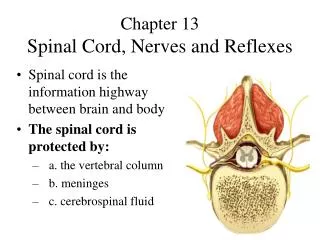
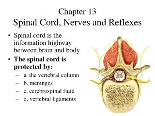
Stretch Reflex • Sudden stretch to a muscle leads to contraction of that muscle is known as stretch reflex. • The basic unit of this activity is a reflex arc comprises of; • Sense organ. • Afferent neuron. • Center • Efferent neuron • Effector organ.
Reflex Arc • Receptor • Muscle spindle • Afferent neuron • Sensory Nerve • Center • Spinal Segment • Efferent neuron • Motor Nerve • Effector Organ • Skeletal Muscle
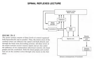
Skeletal Muscle Reflexes • How do they work? • Receptors in muscle send info to CNS • CNS decides should muscle contract (or relax) in response • CNS sends appropriate signal via somatic motor neurons • Somatic motor neurons are always excitatory: • CNS activates if contraction is right response • CNS inhibits if relaxation is right response
Receptors • Muscle receptors: • Sense muscle length and/or tension • Two types of stretch receptors: • Muscle spindles • Golgi tendon organs
Muscle Spindle Structure • Consist of collections of specialized muscle fibers known as intrafusal fibers • Lie within spindle-shaped connective tissue capsules parallel to extrafusal fibers • Each spindle has its own private efferent and afferent nerve supply • Play key role in stretch reflex
Receptor for the stretch reflex: Muscle Spindle Two types of intrafusal fibres: • Nuclear bag fibres: central area is dilated filled with group of nuclei (2 / spindle) • Nuclear chain fibres: smaller than nuclear bag fibres and have one line of nuclei spread in a chain along the receptor area (3-9 /spindle)
Golgi Tendon Organs • Composed of: • Nerve fiber endings that wind between collagen fibers inside connective tissue capsule • If muscle is stretched: • Free nerve endings are pinched and they fire • Activation of Golgi tendon organs: • Inhibits alpha motor neurons and decreases muscle contraction
Stretch Reflex • Primary purpose is to resist tendency for passive stretch of extensor muscles by gravitational forces when person is standing upright • Classic example is patellar tendon, or knee-jerk reflex
The Golgi tendon reflex(inverse stretch reflex) • excessive tension on the muscle (passive stretch of tendon or active muscle contraction) >> muscle relaxes opposite response to stretch reflex. • The receptors are Golgi tendon organs in muscle tendons stimulated >> muscle contract and pulled on the tendon (tension) • stimulate golgi organ>> A fibers > spinal cord > excitation of inhibitory interneuron>> inhibit alpha motor neuron > muscle relaxation • Protect muscle from rupture
Withdrawal reflex(flexor reflex) • Stimulation of pain receptors in hand → impulses to spinal cord via A or C fibres → interneurons →anterior horn cells →stimulate hand flexor muscles → move the hand away from the injurious stimulus. • Its a polysynaptic reflex. • Stimulation of flexors muscle accompanied by inhibition of extensors. • Inhibitory inter neurons synapse with extensor motor neurons known as reciprocal innervations (reciprocal inhibition).
Fig 13.8 – Flexion reflex and the crossed extensor reflex Silverthorn 2nd Ed
Clinical Importance of reflexes • To test the integrity of reflex arc. • Localization of neurological lesion. • Identifying the type of lesion. • Monitoring the progress of neurological deficit.
Clinical Importance of reflexes • Deep tendon reflexes are absent in lower motor neuron lesion. • The become exaggerated in upper motor neuron lesions. • Pendular jerks are observed in cerebellar lesions.