Download
1 / 27
290 likes | 572 Views
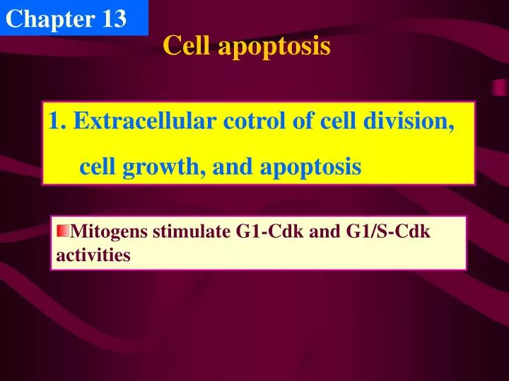
Chapter 13. Cell apoptosis . Extracellular cotrol of cell division, cell growth, and apoptosis. Mitogens stimulate G1-Cdk and G1/S-Cdk activities. 1090 –131= 959(cells). PRIZED Horvitz, and Sulston share Physiology or Medicine Nobel ( 2002 )

E N D
Chapter 13 Cell apoptosis • Extracellular cotrol of cell division, • cell growth, and apoptosis • Mitogens stimulate G1-Cdk and G1/S-Cdk activities
1090 –131= 959(cells) PRIZEDHorvitz, and Sulston share Physiology or Medicine Nobel (2002) “for their discoveries concerning genetic regulation of organ development and programmed cell death”
衰老 机体结构 细胞增殖 细胞分化 细胞凋亡 细胞信号转导 染色体 (DNA与蛋白质的相互作用)
A simplified model of one way that mitogens stimulate cell division
Human cells have a built-in limitation on the number of times they can divide The cell division is cotrolled not only by extracellular mitogens but also by intracellular mechanisms that can limit cell proliferation. Cell-cycle arrest or apoptosis induced by excessive stimulation of mitogenic pathways.
Extracellular growth factors stimulate cell growth Activation of cell-surface receptors Activation of PI 3-kinase Activation of elF4E (translation initiation factor) and S6 kinase(phosphorylates ribosomal protein S6) Increasing protein synthesis and cell growth
2.Apoptosis, Programmed cell death • Biological functions of apoptosis 细胞凋亡是多细胞生物在发育过程中,一种由基因控制的主动的细胞生理性自杀行为。 In development, homeostasis, tumor surveillance, and the function of the immune system.
Morphologi changes: Early : Chromosome condensation, cell body shrink Later : Blebbing and Nucleus and cytoplasm fragment— Apoptotic bodies At last: Phagocytosed • Morphological and biochemical characteristics of apoptosis
A、Normal cell B、Apoptosis: Apoptotic bodies
Biochemical characteristics of apoptosis: 180~200bp DNA ladder, Accumulation of tTG, PS flip-flop Apoptosis induced by Cyto C Lane 1.0 h 2.1 h 3.2 h 4.3 h 5.4 h 6.Control 7.Marker 2.0kbp 1.0 0.5 0.2
Contrast ofApoptosis and necrosis Apoptosis Necrosis Death by apoptosis is a neat, orderly process
3.Molecular mechanisms of apoptosis • Early researches (MIT: Robert Horrid , 1986) C.elegans:1090 cells, The Finding of CED3 mutant Without losing any of their cells to apoptosis. CED3 gene play a crucial role in the process of apoptosis. 131 cells death. C elegans: a millimeter long, transparent body only a few cell types, from zygote to mature adult only in 3.5days.
线虫体细胞凋亡研究:PCD相关基因15个,分为四组:线虫体细胞凋亡研究:PCD相关基因15个,分为四组: • 1、与PCD有关的基因,负责PCD控制:ced-3、ced-4和ced-9。 • 2、与PCD过程吞噬作用有关:ced-1、ced-2、ced-5-8 和 ced-10。 • 3、核酸酶基因(nuc-1), 控制DNA降解,但非PCD所必须。 • 4、影响特异细胞类型PCD的基因:ces-1、ces-2、cgl-1、和 • her-1. • 保守性高,在哺乳动物中有相应同源物: • Ced-3=ICE, Ced-4=Apaf-1, Ced-9=Bcl-2. Mammalian: CED3 is related to mammalian interleukin- 1ß–converting enzyme (ICE or caspase-1)
Apoptosis is carried out by a proteolytic system —caspase (1) Why called caspase? Active site: Cysteine Cleavage site: Asparatic acid Cysteine Asparatic acid specific protease Aps-Xxx 天冬氨酸特异性的半光氨酸蛋白水解酶
Apoptosis can be divided into two phases: Activation phase: The cell responds to “death signals” that commit it to undergoing self-destruction. Execution phase: The death sentence is carried out. Apoptosis cells are recognized by phagocytes because they carry exposed markers, called “eat me” signals. The best studied “eat me” signal is the presence of phosphatidylserine molecules in the outer leaflet of PM of apoptotic cells (by flop-flipase).
All caspases expressed as proenzymes— Procaspases NH2-terminal prodomain: Highly variable • How to activate caspases? Procaspase Large subunit (20kD) Small subunit (10kD) How are procaspases activated to initiate the caspase cascade? The activation is triggered by adaptor proteins that bring multiple copies of specific procaspases. 3 groups of caspase: 1、apoptotic initiators: caspase-2, caspase-8, caspase-9 and caspase-10 2、apoptotic executioners: caspase-3, caspase-6 ,caspase-7 and 14 (morphology change) 3、inflammatory mediateors: caspase-1, and caspase-11
Procaspases are activated by binding to adaptor proteins • The caspase cascade involved in apoptosis • Procaspase activation by proteolytic cleavage. • Caspase cascade
More than a dozen protein kinase, including FAK, PKC, and Raf1. FAK– disrupt cell adhesion for the apoptotic cell. Lamins. Cleavage of lamins leads to the disassembly of the nuclear lamina and shrinkage of the nucleus. Proteins required for cell structure. Such as IF, actin, and gelsilin. Cleavage and inactivation of these proteins lead to changes in cell shape. Induce cell display signals marked it for phagocytosis . • The target proteins of caspase are the following:
The inhibitor of CAD (Caspase-activated Dnase, an endonuclease). Cleavage of CADinhibitor lead to activation of CAD, once activated, CAD translocates from the cytosol to the nucleus severing DNA into fragments. Enzymes involved in DNA repair. Which are inactivated by caspase cleavage. DNA repair is a homeostatic activity that is inappropriate in an apoptotic cell.
Extrinsic pathway Two principle pathways Intrinsic pathway • Molecular pathways of apoptosis
(1)Extrinsic pathway: Fas Signaling Pathway Receptor-mediated pathway of apoptosis Fas (also called Apo-1 or CD95) is a member of the tumor necrosis factor receptor (TNFR) superfamily. Bcl-2 Family(cytoplasmic factors): Bad,Bid,and bax: promote apoptosis; Bcl-X, Bcl-w,and Bcl-2: prevent apoptosis. Internal stimuli: DNA damage, high Ca2+ , Oxidative stress
(2) Intrinsic pathway: Mitochondrial pathway The mitochondria-mediated pathway of apoptosis Various types ofcellular stress Bcl-2 family: Bad or Bax to become inserted into OM of Mit Release of cytochrome c from I-O space of Mit. Form a multisubunit complex; and Caspase Cascade,…
Activation of caspase-2 is required for permeabilization of mitochondria, release of cytochrome c, and apoptosis