Download
1 / 13
150 likes | 326 Views
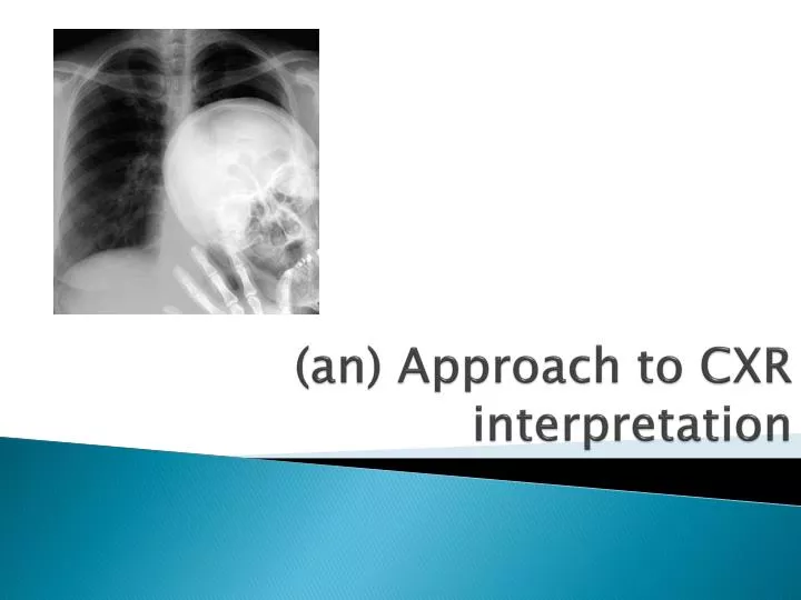
(an) Approach to CXR interpretation. Dr ABCDE’s. Details RIPE image Airway & mediastinum Breathing Circulation Diagphragm Extras Soft tissues & bones. Details. Patient name, age / DOB, sex Type of film – PA or AP, lateral, erect or supine, inspiration or expiration, correct L/R marker

E N D
Dr ABCDE’s • Details • RIPEimage • Airway & mediastinum • Breathing • Circulation • Diagphragm • Extras • Soft tissues & bones
Details • Patient name, age / DOB, sex • Type of film – PA or AP, lateral, erect or supine, inspiration or expiration, correct L/R marker • Date and time of study
RIPE image • Rotation – medial clavicle ends equidistant from spinous process, angulation. • Inspiration – 8-10 posterior ribs above diaphragm, 5-6 anterior ribs. • Picture – straight vs oblique, entire lung fields, scapulae outside lung fields. • Exposure (Penetration) – IV disc spaces and L) hemidiaphragm visible through cardiac shadow
Airway & mediastinum • Trachea – central or slightly to R) as crosses aortic arch • Paratracheal/mediastinal masses or adenopathy • Carina & RMB/LMB • Mediastinal width - <8cm on PA film • Aortic knob • Hilum – L) higher than R) (up to 2-3cm), vessels, calcification
Breathing • Lung fields • Vascularity – to ~2cm of pleural surface (~3cm in apices), vessels in bases > apices • Pneumothorax – don’t forget apices • Lung field outlines – abnormal opacity / lucency, atelectasis, collapse, consolidation, bullae • Horizontal fissure on R) • Pulmonary infiltrates – interstitial vs alveolar pattern • Coin lesions • Cavitary lesions • Pleura • Pleural reflections • Pleural thickening
Circulation • Heart position –⅔ to left, ⅓ to right • Heart size – measure cardiothoracic ratio on PA film (normal <0.5) • Heart borders – R) border is R) atrium, L) border is L) ventricle & atrium • Heart shape • Aortic stripe
Diaphragm • Hemidiaphragm levels – R) higher than L) (~2.5cm / 1 intercostal space) • Diaphragm shape/contour • Cardiophrenic and costophrenic angles – clear and sharp • Gastric bubble / colonic air • Subdiaphragmatic air (pneumoperitoneum)
Extras • ETT, CVP line, NGT, PA catheters, ECG electrodes, PICC line, chest tube • PPM, AIDC, metalwork
Soft tissues and bones • Ribs, sternum, spine, clavicles – symmetry, fractures, dislocations, lytic lesions, density • Soft tissues – looking for symmetry, swelling, loss of tissue planes, subcutaneous air, masses • Breast shadows • Calcification – great vessels, carotids