Download
1 / 43
430 likes | 599 Views
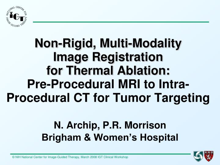
Non-Rigid, Multi-Modality Image Registration for Thermal Ablation: Pre-Procedural MRI to Intra-Procedural CT for Tumor Targeting. N. Archip, P.R. Morrison Brigham & Women’s Hospital. WORKSHOP OBJECTIVE.

E N D
Non-Rigid, Multi-Modality Image Registration for Thermal Ablation:Pre-Procedural MRI to Intra-Procedural CT for Tumor Targeting N. Archip, P.R. Morrison Brigham & Women’s Hospital
WORKSHOP OBJECTIVE To illustrate the need for image registration to address limitations in targeting a tumor during a tumor ablation procedure.
ABLATION GUIDANCE MODALITY ABLATIVE AGENT TREATMENT SITE MRI CRYOTHERAPY LIVER KIDNEY/ADRENAL CT RFA BONE/SOFT TISSUE US ETOH LUNG Acad Radiol 2005;12:1100-109
THERMAL ABLATION • RFA: RADIOFREQUENCY ABLATION • CRYO: CRYOTHERAPY • LITT, ILT: LASER ABLATION • MWA: MICROWAVE ABLATION • HIFU, FUS: FOCUSED ULTRASOUND Images courtesy Cross-sectional Interventional Radiology, Brigham & Women’s Hospital, Boston, MA
CLINICAL SYSTEMS PERCUTANEOUS ABLATION • RF generator & electrode • CRYO unit & probe • LASER & optical fiber • MW generator & antenna Images courtesy Cross-sectional Interventional Radiology, Brigham & Women’s Hospital, Boston, MA
CLINICAL ENIRONMENT Images courtesy Cross-sectional Interventional Radiology, Brigham & Women’s Hospital, Boston, MA
IMAGE-GUIDANCE • DEFINE • PLAN • TARGET • MONITOR • CONTROL • ASSESS Radiology 2005; 236:716-724
DYNAMIC PROCESSES • MRI-guided cryotherapy of a 3.4cm liver metastasis • Sagittal T2-weighted fse imaging used to place cryoprobes (arrowheads) into the tumor (arrow) • Iceballs form around each probe; these coalesce • Tumor is eclipsed Acad Radiol 2005;12:1100-109
3-D PROCESS Pre Intra Post in 3-D Post Modified from AJR 2004;183:707–712
TARGET CT-GUIDED RFA IN LUNG Post-RFA MRI Coronal RFA Images courtesy Cross-sectional Interventional Radiology, Brigham & Women’s Hospital, Boston, MA
PLAN – TX - ASSESS CT-GUIDED RFA IN SOFT TISSUE CT INTRA CE MRI POST CE CT PRE Images courtesy Cross-sectional Interventional Radiology, Brigham & Women’s Hospital, Boston, MA
Non-Rigid, Multi-Modality Image Registration for Thermal Ablation:Pre-Procedural MRI to Intra-Procedural CT for Tumor Targeting
CLINICAL NEED – Example 1 Pre-Procedure CE MRI at 1.5T Intra-Procedure Non-Enhanced CT Recent BWH ablation case: Pre-Proc MRI vs Intra-CT 47yo M, 3cm liver metastasis of sq cell ca in Seg 8; lung & vessels nearby Procedure 071107 Images courtesy Cross-sectional Interventional Radiology, Brigham & Women’s Hospital, Boston, MA
CLINICAL NEED – Example 2 Pre-Procedure CE MRI at 1.5T Intra-Procedure Non-Enhanced CT Recent BWH Ablation Case: PreProc MRI vs Intra-CT 69yo M, 7cm liver metastasis of colon cancer; look for coverage; GB & colon nearby Procedure 080304 Images courtesy Cross-sectional Interventional Radiology, Brigham & Women’s Hospital, Boston, MA
CLINICAL NEED Dx comes from one imaging modality … Tx takes place under another.
SOLUTION: REGISTRATIONBring Pre-procedural diagnostic MRI into the Intra-procedural interventional space
PRE-PROCEDURAL CE MRI Example: Registered Data --MRI Pre BWH Ablation Case 50yo M, 2cm primary liver tumor (HCC) in segment 2 051021 CEMRI PreTx Ser08 i270 Images courtesy Cross-sectional Interventional Radiology, Brigham & Women’s Hospital, Boston, MA
INTRA-PROCEDURAL CT Example: Registered Data --CT Intra 051220 CT PreTx Ser04 i21 Images courtesy Cross-sectional Interventional Radiology, Brigham & Women’s Hospital, Boston, MA
RFA ELECTRODE PLACED Example: Registered Data --CT Intra Images courtesy Cross-sectional Interventional Radiology, Brigham & Women’s Hospital, Boston, MA
REGISTERED IMAGES Example: Registered Data --Merged MRI & CT Images courtesy Cross-sectional Interventional Radiology, Brigham & Women’s Hospital, Boston, MA
WORK FLOW Intra-Proc CT Pre-Proc MRI Registered CT & MRI Images courtesy Cross-sectional Interventional Radiology, Brigham & Women’s Hospital, Boston, MA
WHAT IS 3D SLICER? • A platform for exploring novel image analysis and visualization techniques • A freely-downloadable program • Source code and executables available for Windows, Linux, and Mac OS X • Slicer is a research platform: • NOT an FDA approved medical device • NOT finished – some parts will work better than others
SLICER FEATURES • Visualization • Filtering • Registration • Segmentation • DTI • Quantification • Real-time Integration
“SLICER 3” • Next Generation of the 3D Slicer software • Beta Release in Jan 2007 • Slicer 3.0 schedule for June 2007 • www.slicer.org Image: S Pieper
WHAT IS SLICER 3? • Total Rework of 3D Slicer • At least 80% of code rewritten • New versions of VTK (5.0), ITK (3.2) • Improved Look and Feel (KWWidgets) • Improved Modularity • Analysis routines can be used as plugins or command line executables for batch processing • Draws on Multi-Institution Community • Kitware, Inc., GE Research, Isomics, Inc., BWH, UCLA, UCSD, UIowa, MIT, GaTech, UNC, U Utah, MGH, UCI, Harvard, WashU… • NAC, NCIGT, NA-MIC, mBIRN, fBIRN
SLICER 3 • Open Source, multi-platform • Over 500k lines of code • www.slicer.org • Google: slicer 101 Images: A Golby, F Talos, P Black
Image/Scene Management • XML-Based MRML File Stores Scene Description • Volumes (Images, Label Maps) • Models • Hierarchical Affine Transforms • Scene Data (Cameras, Colors, Fiducials, etc). • Manipulated in World Coordinates based on Patient RAS Image: S Pieper
OPENING SCREEN The default display provides for primary 2D planes and a 3D Viewer space. Other display options can be selected. screen_01
LOAD VOLUME FILE Having selected the Volumes Module … Images can be selected as DICOM or .nrrd files. Here, a pre-procedural MRI scan of the liver is opened. screen_03
VISUALIZE MRI DATA MRI opens in three orthogonal 2D planes. screen_04
TUMOR LOCATION 3D image planes can be viewed in the 3D space. Can zoom, translate, rotate. Here we locate tumor in all 3 planes (arrows) screen_05
3-D ONLY View MRI planes in 3D Viewer from above … and from below. screen_06 & 07
Add Volume CT Add a volume to the space … Here, an intra-procedural CT dataset is opened. screen_08
VISUALIZE CT DATA CT opens in three orthogonal 2D planes. screen_09
PREP FOR LINEAR TRANSFORM Initial alignment of MRI to CT is the Linear Transform 1 under Transform Module. This manual transform is one of two options. screen_10
LINEAR TRANSFORM Initial linear alignment solves initial displacement of MRI and CT. Seen here in the 3D Viewer. screen_20
PREP FOR AUTO TRANSFORM Initial alignment of MRI to CT can be done by selecting Linear under Registration Module menu. This automatic transform is the second of two options. screen_22
RESULT OF RIGID REGISTRATION Slider can be used to review the result of the linear alignment between the pre-procedural MRI and intra-procedural CT. screen_21c
PREP FOR B-SPLINE REGISTRATION Alignment can be done on the data using the Deformable B-Spline B-Spline Registration option. screen_25
RESULT OF B-SPLINE REGISTRATION Slider can be used to review the result of the linear alignment between the pre-procedural MRI and intra-procedural CT. screen_26
CLINICAL SIGNIFICANCE Image Registration has relevance for all aspects of an ablation procedure: Plan, Target, Monitor, Control, AssessThis is true for any image guidance modality.