Download

1 / 28
350 likes | 723 Views
Metabolic Oxidative Stress and Cancer Douglas R. Spitz, PhD Free Radical and Radiation Biology Program Department of Radiation Oncology Holden Comprehensive Cancer Center The University of Iowa, Iowa City, IA. What is life?

E N D
Metabolic Oxidative Stress and Cancer Douglas R. Spitz, PhD Free Radical and Radiation Biology Program Department of Radiation Oncology Holden Comprehensive Cancer Center The University of Iowa, Iowa City, IA
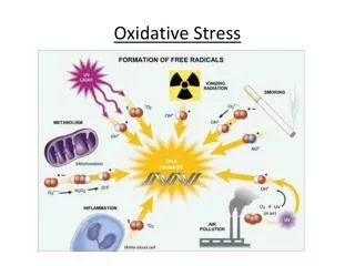
What is life? “An electron, given off easily, has a high energy level, while an orbital which tends to take up an electron, has a low energy level. The transmitted electron thus goes from a high to a low level, and releases the energy which corresponds to the difference of the two levels. It is this energy which drives life.” Albert Szent-Györgyi, Electronic Biology and Cancer, Marcel Dekker Inc. 1976 In essence, Szent-Györgyi was one of the first scientists to recognize that all of the forces necessary for the maintenance of living systems derive from the ability of complex higher order biological structures to extract, store, and move electrons.
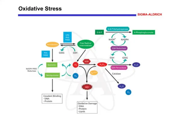
Why do we breath oxygen? The Chemiosmotic Hypothesis Why is it potentially harmful? Lehninger; Principles in Biochemistry 3 rd Edition Chemiosmotic model. In this simple representation of the chemiosmotic theory applied to mitochondria, electrons from NADH and other oxidizable substrates pass through a chain of carriers arranged asymmetrically in the inner membrane. Electron flow is accompanied by proton transfer across the membrane, producing both a chemical gradient (pH) and an electrical gradient (). The inner mitrochondrial membrane is impermeable to protons; protons can reenter the matrix only through proton specific channels (F0). The proton-motive force that drives protons back into the matrix provides the energy for ATP synthesis, catalyzed by the F1 complex associated with F0.
Ahmad IM, Aykin-Burns N, Sim JE, Walsh SA, Higashikubo R, Buettner GR, Venkataraman S, Mackey MA, Flanagan S, Oberley LW, and Spitz DR: Mitochondrial O2- and H2O2 mediate glucose deprivation-induced cytotoxicity and oxidative stress in human cancer cells. J. Biol. Chem. 2005; 280(6):4254-4263.
Metabolic Theories of Cancer Warburg O (1956) Science 123:309-314 Cancer cells exhibit increased rates of glycolysis and slightly decreased rates of respiration. It was proposed this was the result of “damage” to the respiratory mechanism and tumor cells increased glycolysis to compensate for this defect. Weber G (1977) New Engl. J. Med. 296:541-551 Cancer cells exhibit increased rates of pentose phosphate cycle activity characterized by increases in glucose-6-phosphate dehydrogenase activity. Oberley LW, Oberley TD, and Buettner GR (1980 and 1981) Med. Hypoth. 6:49-68; 7:21-42 Tumor cells have aberrant respiration caused by a decrease in MnSOD activity leading to increased steady state levels of superoxide and hydrogen peroxide that causes DNA damage and activates signaling pathways leading to uncontrolled growth, the inability to differentiate, and the malignant phenotype.
Genetic Theory of Cancer Bishop (1987) Science 235:305-311; Varmus (1987) Science 238:1337-1339 Cancer is a multi-step genetic disease in which mutations resulting in the aberrant expression of cellular homologues of oncogenes (i.e., Ras, c-Fos, c-Jun, and c-Myc, etc.) associated with growth and development as well as tumor suppressor genes (i.e., p53) gradually accumulate over time, eventually resulting in immortalization, the loss of control of cell proliferation, and progression to the malignant phenotype. How can we unify metabolic and genetic theories of cancer?
In the mid-1990’s Dr. Yong J. Lee’s laboratory showed: Within minutes - glucose deprivation-induced activation of signal transduction in (i.e., ERK1, ERK2, Lyn Kinase, JNK, Ras, AP-1, etc.) in MCF-7/ADR human breast cancer cells. Within 2-4 hours - glucose deprivation-induced increases in steady state levels of mRNA coding for bFGF and c-Myc in MCF-7/ADR. Within 4 hours - glucose deprivation-induced clonogenic cell killing in MCF-7/ADR cells. Molecular and Cellular Biochemistry, 155:163-171, 1996. Journal of Cell Science, 110:681-686, 1997. Molecular and Cellular Biochemistry, 170:23-30, 1997. Journal of Biological Chemistry, 272:11690-11693, 1997. This work supported the existence of a mechanistic link between glucose metabolism, signal transduction, and gene expression governing the malignant phenotype that also governed the survival of cancer cells. What is this mechanistic link?
Dr. Lee and his collaborators then discovered that all the effects of glucose deprivation in MCF-7 ADR could be suppressed by treatment with the thiol antioxidant, N-acetylcysteine, indicating a causal role for oxidative stress in the effects of glucose deprivation in cancer cells. Lee YJ, Galoforo SS, Berns CM, Chen JC, Davis BH, Sim JE, Corry PM, and Spitz DR: Glucose deprivation-induced cytotoxicity and alterations in mitogen-activated protein kinase activation are mediated by oxidative stress in multidrug- resistant human breast carcinoma cells.J. Biol. Chem.1998; 273:5294-5299. Blackburn RV, Spitz DR, Liu X, Galoforo SS, Sim JE, Ridnour LA, Chen JC, Davis BH, Corry PM, and Lee YJ: Metabolic oxidative stress activates signal transduction and gene expression during glucose deprivation in human tumor cells.Free Radic. Biol. Med.1999; 26:419-430.
Kinases (ie, Lyn, PKC, etc.) G proteins (ie, Ras, etc.) DNA Kinases (ie, Raf, MEK, ERK, JNK, etc.) Phosphatases RNA (ie, c-Fos, Trxn. factors (ie, AP-1, etc.) c-Jun, c-Myc, bFGF, etc.) Glucose ? NUCLEUS PPC NADP+ GSH H O 2 2 Glucose 6-phosphate Proteins Glycolysis GR GPX H O 2 2 NADPH GSSG H O Pyruvate 2 O H O • 2 2 2 H O 1 e- Site II 2 Site III Site I Site IV NADH Fatty/Amino 4e- TCA Acids H O 2 FADH 2 O 2 MITOCHONDRIA PLASMA MEMBRANE Theoretical model outlining a biochemical rationale for unifying metabolic and genetic theories of cancer. Spitz et al. (2000) Ann. N.Y. Acad. Sci. 899:349-362. ? = thioredoxin, glutaredoxin, and direct redox changes on proteins (TrxS2H2) (TrxPx) (TR) (TrxSS)
Rodent tumor cells had reduced levels of MnSOD activity that were proportional to increased growth rate and lack of ability to differentiate. Rodent Tumor cells demonstrated mitochondrial abnormalities that were more pronounced in fast growing undifferentiated cancers. Bize IB, Oberley LW, Morris HP (1980) Superoxide dismutase and superoxide radical in Morris hepatomas. Cancer Res. 40(10):3686-93. normal liver well differentiated undifferentiated mitochondria hepatoma hepatoma slow growing fast growing
Human cancer cells demonstrated mitochondrial abnormalities that are more pronounced in more malignant tissues. Springer EL: Comparative study of the cytoplasmic organelles of epithelial cell lines derived from human carcinomas and nonmalignant tissues. Cancer Res. 1980; 40(3):803-17. The cytoplasmic organelles of 16 human cell types derived from normal as well as from primary and metastatic carcinomas were characterized by electron microscopy in a blinded fashion. Mitochondrial pleomorphisms was expressed slightly by normal cells and to a much greater extent by all cell types derived from malignant tissues. These pleomorphisms were characterized by hypertrophied mitochondria with longitudinal cristal arrangements in almost all the malignant cells lines, but not in any lines derived from nonmalignant tissues of cancerous organs or from normal tissues.
Increasing mitochondrial superoxide scavenging by increasing expression of MnSOD alters the malignant phenotype of cancer cells Church SL, Grant JW, Ridnour LA, Oberley LW, Swanson PE, Meltzer PS, Trent JM: Increased manganese superoxide dismutase expression suppresses the malignant phenotype of human melanoma cells.Proc Natl Acad Sci U S A. 1993; 90(7):3113-7. Melanoma cell lines over expressing MnSOD cDNAs lost their ability to form colonies in soft agar and tumors in nude mice. These findings were the first demonstration that stable over expression MnSOD suppressed the malignant phenotype in human cancer cells These results suggested that increased steady-state levels of superoxide and/or H2O2 significantly contributed to the maintenance of the malignant phenotype in human cancer cells.
Human cancer cells were reported to produce large amounts of H2O2, relative to normal cells Szatrowski TP and Nathan CF: Production of large amounts of hydrogen peroxide by human tumor cells. Cancer Res. 1991; 51(3):794-8. Seven human tumor cell lines demonstrated constitutively elevated H2O2 such that cumulative amounts were comparable to the amount of H2O2 produced by phorbol ester-activated neutrophils. Constitutive generation of large amounts of reactive oxygen intermediates by cancer cells, if it occurs in vivo, might contribute to genomic instability, tumor heterogeneity, invasion, and metastasis.
Human colon carcinoma cells demonstrate increased steady-state levels of superoxide relative to normal human colon epithelial cells and fibroblasts Mitochondrial electron transport chain blockers make this difference more pronounced • Aykin-Burns N, Ahmad IM, Zhu Y, Oberley LW, and Spitz DR: Inhibition of glucose metabolism causes enhanced cytotoxicity in human cancer vs. normal cells via metabolic oxidative stress. Biochem. J. 2009; 418:29-37.
Human breast carcinoma cells demonstrate increased steady-state levels of superoxide and hydroperoxides, relative to normal human breast epithelial cells • Aykin-Burns N, Ahmad IM, Zhu Y, Oberley LW, and Spitz DR: Inhibition of glucose metabolism causes enhanced cytotoxicity in human cancer vs. normal cells via metabolic oxidative stress. Biochem. J. 2009; 418:29-37.
Human colon and breast carcinoma cells demonstrate increased sensitivity to glucose deprivation-induced cytotoxicity, relative to normal human cells • Aykin-Burns N, Ahmad IM, Zhu Y, Oberley LW, and Spitz DR: Inhibition of glucose metabolism causes enhanced cytotoxicity in human cancer vs. normal cells via metabolic oxidative stress. Biochem. J. 2009; 418:29-37.
Mitochondrial O2- and H2O2 mediate glucose deprivation-induced toxicity and oxidative stress in human cancer cells. • Ahmad IM, Aykin-Burns N, Sim JE, Walsh SA, Higashikubo R, Buettner GR, Venkataraman S, Mackey MA, Flanagan S, Oberley LW, and Spitz DR: Mitochondrial O2- and H2O2 mediate glucose deprivation-induced cytotoxicity and oxidative stress in human cancer cells. J. Biol. Chem. 2005; 280(6):4254-4263. • Aykin-Burns N, Ahmad IM, Zhu Y, Oberley LW, and Spitz DR: Inhibition of glucose metabolism causes enhanced cytotoxicity in human cancer vs. normal cells via metabolic oxidative stress. Biochem. J. 2009; 418:29-37.
Human colon carcinoma cells are more sensitive to cell killing by 2-deoxyglucose (2DG), relative to normal colon epithelial cells 2DG-induced cytotoxicity can be inhibited by over expressing mitochondrially targeted catalase and superoxide dismutase • Aykin-Burns N, Ahmad IM, Zhu Y, Oberley LW, and Spitz DR: Inhibition of glucose metabolism causes enhanced cytotoxicity in human cancer vs. normal cells via metabolic oxidative stress. Biochem. J. 2009; 418:29-37
Mutations in mtDNA encoded genes (ND6) can lead to increased ROS and increased metastatic potential that is suppressed by ROS scavengers. Ishikawa K, Takenaga K, Akimoto M, Koshikawa N, Yamaguchi A, Imanishi H, Nakada K, Honma Y, Hayashi J: ROS-generating mitochondrial DNA mutations can regulate tumor cell metastasis. Science 2008; 320(5876):661-4. Cytoplasmic hybrid (cybrid) technology was used to replace endogenous mtDNA in a mouse tumor cell line that was poorly metastatic with mtDNA from a highly metastatic cell line and the recipient tumor cells acquired the metastatic potential of the transferred mtDNA. The mtDNA conferring high metastatic potential contained mutations in the gene encoding NADH (reduced form of nicotinamide adenine dinucleotide) dehydrogenase subunit 6 (ND6) that was associated with overproduction of reactive oxygen species (ROS). Pretreatment of the highly metastatic tumor cells with ROS scavengers suppressed their metastatic potential in mice.
Mutations in nuclear encoded genes that code for electron transport chain proteins (SDH subunits B, C, D) can also lead to increased steady-state levels of superoxide, increased mutation rates, increased genomic instability, and tumorigenesis in rodents as well as being associated with neuro-endocrine tumor formation in humans. Ishii T, Yasuda K, Akatsuka A, Hino O, Hartman PS, Ishii N: A mutation in the SDHC gene of complex II increases oxidative stress, resulting in apoptosis and tumorigenesis. Cancer Res 2005; 65(1):203-9. Slane BG, Aykin-Burns N, Smith BJ, Kalen AL, Goswami PC, Domann FE, Spitz DR: Mutation of succinate dehydrogenase subunit C results in increased O2-, oxidative stress, and genomic instability. Cancer Res 2006; 66(15):7615-20. Bardella C, Pollard PJ, Tomlinson I: SDH mutations in cancer. Biochim Biophys Acta 2011; 1807(11):1432-43. Owens KM, Aykin-Burns N, Dayal D, Coleman MC, Domann FE, and Spitz DR: Genomic instability induced by mutant succinate dehydrogenase subunit D (SDHD) is mediated by O2- and H2O2. Free Radic Biol Med 2011; [Epub ahead of print] PMID: 22041456
Conclusions and Speculations: Cancer cells appear to exist in a condition of metabolic oxidative stress characterized by increased steady-state levels of superoxide and hydrogen peroxide. This increase in reactive oxygen species (ROS) appears to be compensated for by increases in glucose and hydroperoxide metabolism. The condition of metabolic oxidative stress in cancer cells may be the result of alterations in mitochondrial oxidative metabolism. Metabolic oxidative stress appears to contribute to selective sensitivity of cancer vs. normal cells to glucose deprivation-induced cytotoxicity and oxidative stress.
SPECULATION • Cancer might represent a constellation of metabolic and/or genetic diseases where the common theme is uncoupling of normal cellular processes that govern cell growth and differentiation caused by the inappropriate flow of electrons from metabolic oxidation/reduction reactions to redox sensitive proteins governing signal transduction and gene expression. (Spitz, et al. Ann. N.Y. Acad. Sci. 899:349-362, 2000)
Clinical Implications: Therapy If glucose metabolism is increased in cancer cells to compensate for excess hydroperoxide production from mitochondrial respiration, then inhibiting glucose and hydroperoxide metabolism while forcing cells to derive energy from respiration should preferentially kill cancer cells, relative to normal cells. Combinations of agents designed to take advantage of this hypothesis might include: Inhibitors of Glucose Metabolism (ie., 2-deoxyglucose, etc.) Inhibitors of the Pentose Phosphate Cycle (ie., DHEA, etc.) Inhibitors of Hydroperoxide Metabolism (ie., BSO, AUR, BCNU) Dietary Manipulation (high protein/fats, no carbohydrates) Agents that Increase the Metabolic Production of Prooxidants and Increase Oxidative DNA Damage (ie., Radiation, quinones, Cisplatin, Paclitaxel, inflammatory mediators, etc.)
Inhibitors of glucose and hydroperoxide metabolism selectively (relative to normal cells) enhance cancer cell killing as well as selectively sensitizing cancer cells to therapeutic agents. • Lin X, Zhang F, Bradbury CW, Kaushal A, Li L, Spitz DR, Aft R, and Gius D: 2-Deoxy-d-glucose-induced cytotoxicity and radiosensitization in tumor cells is mediated via disruptions in thiol metabolism. Cancer Res 2003; 63:3413–3417. • Simons AL, Ahmad IM, Mattson DM, Dornfeld KJ, and Spitz DR: 2-Deoxy-D-glucose combined with cisplatin enhances cytotoxicity via metabolic oxidative stress in human head and neck cancer cells. Cancer Res 2007; 67(7): 3364–70. • Coleman MC, Asbury CR, Daniels D, Du J, Aykin-Burns N, Smith BJ, Li L, Spitz DR, Cullen JJ: 2-deoxy-D-glucose causes cytotoxicity, oxidative stress, and radiosensitization in pancreatic cancer. Free Radic Biol Med 2008; 44(3):322-31. • Fath MA, Diers AR, Aykin-Burns N, Simons AL, Hua L, and Spitz DR: Mitochondrial electron transport chain blockers enhance 2-deoxy-D-glucose induced oxidative stress and cell killing in human colon carcinoma cells. Cancer Biol Ther 2009; 8(13):1228-36. • Hadzic T, Aykin-Burns N, Coleman MC, Zhu Y, Jacobson G, and Spitz DR: Paclitaxel combined with inhibitors of glucose and hydroperoxide metabolism enhances breast cancer cell killing via H2O2-mediated oxidative stress. Free Radic Biol Med 2010; 48(8):1024-33. (in press). • Sinthupibulyakit C, Ittarat W, St Clair WH, St Clair DK: p53 Protects lung cancer cells against metabolic stress. Int J Oncol. 2010; 37(6):1575-81. • Shutt DC, O'Dorisio MS, Aykin-Burns N, Spitz DR: 2-deoxy-D-glucose induces oxidative stress and cell killing in human neuroblastoma cells. Cancer Biol Ther 2010; 9(11):853-61. • Sharma PK, Bhardwaj R, Dwarakanath BS, Varshney R: Metabolic oxidative stress induced by a combination of 2-DG and 6-AN enhances radiation damage selectively in malignant cells via non-coordinated expression of antioxidant enzymes. Cancer Lett. 2010; 295(2):154-66. • Fath M, Ahmad IM, Smith CJ, Spence JM, Spitz DR: Enhancement of carboplatin-mediated lung cancer cell killing by simultaneous disruption of glutathione and thioredoxin metabolism. Clin Cancer Res. 2011; [Epub ahead of print] • Scarbrough PM, Mattson DM, Gius D, Watson WH, and Spitz DR: Simultaneous inhibition of glutathione- and thioredoxin-dependent metabolism is necessary to potentiate 17AAG-induced cancer cell killing via oxidative stress. Free Radic Biol Med 2011; (in press).
Clinical Implications: Imaging • If glucose metabolism is increased in cancer cells to compensate for increased production of prooxidants by mitochondrial metabolism, then alterations in glucose metabolism and mitochondrial function should be proportional to susceptibility to therapies based on taking advantage of this metabolic defect to selectively kill cancer cells. • Use FDG-PET imaging to correlate changes in glucose metabolism (FDG uptake) with sensitivity to: • Radiation + 2DG • Cisplatin + 2DG • Simons AL, Fath MA, Mattson DM, Smith BJ, Walsh SA, Graham MM, Hichwa RD, Buatti JM, Dornfeld KJ, and Spitz DR: Enhanced response of human head and neck cancer xenograft tumors to Cisplatin combined with 2-deoxy-D-glucose correlates with increased 18F-FDG uptake as determined by PET imaging. Int J Radiat Oncol Biol Phys 2007; 69(4):1222-1230.
Conclusions and Speculations: Metabolic oxidative stress appears to contribute to the selective sensitization of cancer vs. normal cells to 2DG and inhibitors of glutathione and thioredoxin metabolism in a variety of human cancer cell models. Inhibition of glucose and hydroperoxide metabolism may provide a biochemical target for selectively enhancing cytotoxicity and oxidative stress in cancer cells treated with conventional therapeutic agents (radiation and/or chemotherapy) that cause oxidative stress in clinical trials. Taking advantage of fundamental differences in oxidative metabolism between cancer vs. normal cells to design combined modality cancer therapies, could provide improve outcomes while limiting normal tissue injury by allowing for the de-escalation of doses of highly cytotoxic agents while maintaining or improving efficacy of cancer cell killing.
Thanks for your attention! Supported by: R01CA133114; R21CA139182; R21CA161182; T32CA078586; P30CA086862