Download
1 / 26
290 likes | 854 Views
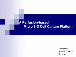
Alginate Sponges For 3-D Cell Culture. Smadar Cohen & Tsiona Elkayam Ben-Gurion University of the Negev. Alginate Sponge Characteristics. Alginate Sponge 90% Porosity; ~200 m m Pore Size. 200 m m. Cell Distribution within Alginate Sponge (10mm thick). H&E cross sections.

E N D
Alginate SpongesFor 3-D Cell Culture Smadar Cohen & Tsiona Elkayam Ben-Gurion University of the Negev Cohen_Ben-Gurion University
Alginate Sponge Characteristics Cohen_Ben-Gurion University
Alginate Sponge90% Porosity; ~200mm Pore Size 200mm Cohen_Ben-Gurion University
Cell Distribution within Alginate Sponge (10mm thick) H&E cross sections Initial cell seeding density (1x10⁸ cells/cm3) Surface MTT Center SEM Cohen_Ben-Gurion University Bottom
3-D Culture in Alginate Sponge Cohen_Ben-Gurion University
3-D Cell Applications with Alginate Sponges • Tissue engineering • Bioproduction of valuable therapeutics • ADMETox research • Expansion of hematopoeitic and mesenchymal stem cells • Stem cell differentiation Cohen_Ben-Gurion University
Tissue Engineering of Cardiac Muscle Cohen_Ben-Gurion University Schwarzkopf et al, Tissue Engineering, 12 (2006)
Organization of Primary Hepatocytes as Spheroids within Alginate Sponges Enhanced Hepatocellular Functions FDA staining SEM TEM Glicklis et al (2000, 2004); Dvir-Ginzberg et al (2003, 2004); Cohen_Ben-Gurion University
Alginate Sponges for 3-D Stem Cells • The sponge recapitulates the stem cellniche 3-D microenvironment • The scaffold can be made cell specific by attaching adhesion peptides (RGD, YIGSR) • The scaffold can be made a reservoirfor growth factors and molecular agents important for cell growth and differentiation • High cell density can be reachedin the scaffold, without reaching confluence Cohen_Ben-Gurion University
Stem Cell R&D in Alginate Scaffold • Expansion and a better control over differentiationof Human Embryonic Stem (hES) Cells • Expansion of Hematopoietic stem cells (HSC) • Expansion and induced differentiation of Mesenchymal stem cells (MSC) Cohen_Ben-Gurion University
Human Embryonic Stem Cells (hESC) Alginate sponge allows colonization of undifferentiated hESC within pores Alginate sponge pore sizecontrols the aggregationof human embryoid bodies (hEB) The alginate sponge maximizes hES cell expansion and has a better control over cell differentiation Cohen_Ben-Gurion University
Morphology of hEBs Petri-Dish Extensive Aggregation & Central Necrosis Scaffold-borne hEBs Homogenous and Small Size No Central Necrosis Gerecht-Nir et al, Biotechnol. Bioeng. (2004) Cohen_Ben-Gurion University
Key Features of Sponge-borne hEBs • hEBs ranging between 250-900 mm after 1 month, smaller than hEBs formed in conventional Petri dishes • Spherical, homogenous in size relative to those formed in Petri dishes • NO CENTRAL NECROSIS in hEBs formed in alginate sponges Cohen_Ben-Gurion University
Expansion of Human Embryonic Stem Cells (hESC) in Alginate Sponges Seeding: 0.1-1 x10⁵/scaffold Alginate Sponge Petri Dish 4-Fold higher expansion in scaffold-borne EBs relative to Petri-dish Cohen_Ben-Gurion University
ECTODERM ENDODERM MESODERM Differentiation within Alginate Borne-hEBsinto 3 Germ Layers H&E Hepatocyte-like cells (a-FP+) Neuronal cells (nestin+) Cohen_Ben-Gurion University
Case Story: Alginate vs. Collagen Scaffolds for ADMET Collagen Alginate Cohen_Ben-Gurion University
C3A- Clone Derivative of HepG2 Cohen_Ben-Gurion University
C3A Cell Morphology in Alginate vs. Collagen Scaffolds 3-D Spheroids 2-D Adherent Cells Collagen Alginate Cohen_Ben-Gurion University
Laminin Expression in C3A seeded in Alginate vs. Collagen Scaffolds Collagen Alginate Cohen_Ben-Gurion University
C3A Spheroids Express Albumin& CK18 Cohen_Ben-Gurion University
CYP3A4 Metabolism in C3A Spheroids Cohen_Ben-Gurion University
CYP1A2 Metabolism in 2D vs. 3D Cultures Cohen_Ben-Gurion University
1A2 mRNA levels in C3A seeded in Alginate vs. Collagen Scaffolds Cohen_Ben-Gurion University
Summary- ADMET • 3-D culture of C3A in alginate scaffolds induces spheroid formation • Spheroids reveal high levels of hepatocellular functions (protein secretion, drug metabolism and detoxification) • This construct is suitable for HTS initial drug screening (ADMET) Cohen_Ben-Gurion University
Summary- Why Alginate Sponges? • Ready to use for 3-D cell Culture • Algae-derived hydrogel • Optimized for specific cell types • Enhances cell activity • Degrades without changing local pH • Prolonged shelf life • Dimensionally stable in culture Cohen_Ben-Gurion University