Download
1 / 37
380 likes | 573 Views
Humoral Immunity & Immunoglobulin Structure and Function. Dr. Adel Almogren. Humoral ( Antibody-Mediated ) Immunity + Involves production of antibodies against foreign antigens. + Antibodies are produced by a subset of lymphocytes called B cells .

E N D
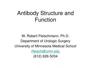
Humoral Immunity &Immunoglobulin Structure and Function Dr. Adel Almogren
Humoral (Antibody-Mediated) Immunity • + Involves production of antibodies against foreign antigens. • + Antibodies are produced by a subset of lymphocytes called B cells. • + B cells that are stimulated will actively secrete antibodies and are called plasma cells. • + Antibodies are found in extracellular fluids (blood plasma, lymph, mucus, etc.) and the surface of B cells. • + Defense against bacteria, bacterial toxins, and viruses that circulate freely in body fluids, before they enter cells. • + Also cause certain reactions against transplanted tissue.
Thymus-independent because T cells are not needed Thymus-dependent because T cells are required Some responses require T help whereas other do not B cell activation T-independent antibody response generally have1. no memory2. no isotype switching3. no somatic mutations
Fab Fc S VL CL S S S CH3 S S CH2 CH1 VH S S F(ab)2 Pepsin cleavage sites - 1 x (Fab)2 & 1 x Fc Papain cleavage sites - 2 x Fab 1 x Fc Domain Structure of Immunoglobulins Domains are folded, compact, protease resistant structures Light chain C domains k or l Heavy chain C domains a, d, e, g, or m
CH2 CH3
CH1 CH2 CH3
VH1 CH1 CH2 CH3
VH1 CH1 CL CH2 CH3
VH1 CH1 VL CL CH2 CH3
VH1 CH1 VL CL CH2 CH3
VH CH1 CL VL CH2 Elbow Hinge CH3
Fv Fv Fv Elbow Fb Fb Fv Fb Fv Hinge CH2 CH2 CH3 CH3 Flexibility and motion of immunoglobulins
FR1 CDR1 FR2 CDR2 FR3 CDR3 FR4 100 Variability 80 60 40 20 20 40 60 80 100 120 Amino acid No. Hypervariable regions • Most hypervariable regions coincided with antigen contact points - the COMPLEMENTARITY DETERMINING REGIONS (CDRs)
Hypervariable CDRs are located on loops at the end of the Fv regions
Hypervariable loops and framework: Summary • The sequences of the hypervariable loops are highly variable amongst antibodies of different specificities • Variable amino acid sequence in the hypervariable loops accounts for the diversity of antigens that can be recognised by a repertoire of antibodies
Electron micrographs of the effect of antibodies and complement upon bacteria Healthy E. coli Antibody + complement- mediated damage to E. coli
IgA IgD IgG CH3 CH2 IgE IgM The hinge region is replaced by an additional Ig domain CH4 CH2 CH3 Structure and function of the Fc region Fc structure is common to all specificities of antibody within an ISOTYPE (although there are allotypes) The structure acts as a receptor for complement proteins and a ligand for cellular binding sites
Cm4 N.B. Only constant heavy chain domains are shown Cm2 Cm3 Cm1 Monomeric IgM IgM only exists as a monomer on the surface of B cells Monomeric IgM has a very low affinity for antigen IgM forms pentamers and hexamers
C C Cm4 Cm4 Cm4 Cm4 Cm4 Cm2 Cm2 Cm2 Cm2 Cm2 Cm3 Cm3 Cm3 Cm3 Cm3 C s s s s s s s s C C C C C C C C C C C C C C C Multimerisation of IgM
IgM facts and figures Heavy chain: m - Mu Half-life: 5 to 10 days % of Ig in serum: 10 Serum level (mgml-1): 0.25 - 3.1 Complement activation:++++ by classical pathway Interactions with cells: Phagocytes via C3b receptors Epithelial cells via polymeric Ig receptor Transplacental transfer: No Affinity for antigen: Monomeric IgM - low affinity - valency of 2 Pentameric IgM - high avidity - valency of 10
IgD facts and figures Heavy chain: d - Delta Half-life: 2 to 8 days % of Ig in serum: 0.2 Serum level (mgml-1): 0.03 - 0.4 Complement activation:No Interactions with cells:T cells via lectin like IgD receptor Transplacental transfer: No ??IgD & IgM ??
S S S S s s C J C S S C C S S C C IgA dimerisation and secretion IgA is the major isotype of antibody secreted at mucosal surfaces Exists in serum as a monomer, but more usually as a J chain-linked dimer, that is formed in a similar manner to IgM pentamers. IgA exists in two subclasses IgA1 is mostly found in serum and made by bone marrow B cells IgA2 is found in higher concentration in mucosal secretions, colostrum
S S S S S S S S S S S S S S S S S S S S IgA and pIgR are transported to the apical surface in vesicles Epithelial cell s s s s s s s s s s C C C C C J J J J J C C C C C S S S S S S S S S S C C C C C C C C C C pIgR & IgA are internalised S S S S S S S S S S Polymeric Ig receptors are expressed on the basolateral surface of epithelial cells to capture IgA produced in the mucosa C C C C C C C C C C B B cells located in the submucosa produce dimeric IgA Secretory IgA and transcytosis ‘Stalk’ of the pIgR is degraded to release IgA containing part of the pIgR - the secretory component
IgA facts and figures Heavy chains:a1or a2 - Alpha 1 or 2 Half-life: IgA1 5 - 7 days IgA2 4 - 6 days Serum levels (mgml-1): IgA1 1.4 - 4.2 IgA2 0.2 - 0.5 % of Ig in serum: IgA1 11 - 14 IgA2 1 - 4 Complement activation:IgA1 - by alternative and lectin pathway IgA2 - No Interactions with cells: Epithelial cells by pIgR Phagocytes by IgA receptor Transplacental transfer: No
IgE facts and figures Heavy chain: e - Epsilon Half-life: 1 - 5 days Serum level (mgml-1):0.0001 - 0.0002 % of Ig in serum: 0.004 Complement activation:No Interactions with cells:Via high affinity IgE receptors expressed by mast cells, eosinophils, basophils and Langerhans cells Via low affinity IgE receptor on B cells and monocytes Transplacental transfer: No its role in protecting against parasitic infections IgE is also closely linked with allergic diseases
IgG facts and figures Heavy chains:g 1 g 2 g3 g4 - Gamma 1 - 4 Half-life: IgG1 21 - 24 days IgG2 21 - 24 days IgG3 7 - 8 days IgG4 21 - 24 days Serum level (mgml-1):IgG1 5 - 12 IgG2 2 - 6 IgG3 0.5 - 1 IgG4 0.2 - 1 % of Ig in serum: IgG1 45 - 53 IgG2 11 - 15 IgG3 3 - 6 IgG4 1 - 4 Complement activation:IgG1 +++ IgG2 + IgG3 ++++ IgG4 No Interactions with cells: All subclasses via IgG receptors on macrophages and phagocytes Transplacental transfer: IgG1 ++ IgG2 + IgG3 ++ IgG4 ++ The neonatal Fcg receptor may be responsible!
C1q binding motif is located on the Cg2 domain Carbohydrate is essential for complement activation Subltly different hinge regions between subclasses accounts for differing abilities to activate complement
Fcg receptors Receptor Cell type Effect of ligation FcgRI Macrophages Neutrophils, Eosinophils, Dendritic cells Uptake, Respiratory burst FcgRIIAMacrophages Neutrophils, Eosinophils, Platelets Langerhans cells Uptake, Granule release FcgRIIB1 B cells, Mast Cells No Uptake, Inhibition of stimulation FcgRIIB2Macrophages Neutrophils, Eosinophils Uptake, Inhibition of stimulation FcgRIIINK cells, Eosinophils, Macrophages, Neutrophils Mast cells Induction of killing (NK cells)
Fv VH1 Fb CH1 VL CL Fab CH2 Elbow Hinge Fc Carbohydrate CH3
Antibody Dependent Cell Mediated Cytotoxicity (ADCC) • Target cell is covered with antibodies, leaving Fc portion sticking outwards. • Natural killer and other nonspecific cells that have receptors for Fc region are stimulated to kill targeted cells. • Target organism is lysed by substances secreted by attacking cells. • Used to destroy large organisms that cannot be phagocytosed.