Download
1 / 114
1.18k likes | 1.55k Views
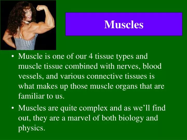
Muscles. Muscle is one of our 4 tissue types and muscle tissue combined with nerves, blood vessels, and various connective tissues is what makes up those muscle organs that are familiar to us. Muscles are quite complex and as we’ll find out, they are a marvel of both biology and physics.

E N D
Muscles • Muscle is one of our 4 tissue types and muscle tissue combined with nerves, blood vessels, and various connective tissues is what makes up those muscle organs that are familiar to us. • Muscles are quite complex and as we’ll find out, they are a marvel of both biology and physics.
Muscle Functions • Production of Movement • Movement of body parts and of the environment • Movement of blood through the heart and the circulatory vessels. • Movement of lymph through the lymphatic vessels • Movement of food (and, subsequently, food waste) through the GI tract • Movement of bile out of the gallbladder and into the digestive tract • Movement of urine through the urinary tract • Movement of semen through the male reproductive tract and female reproductive tract • Movement of a newborn through the birth canal
Muscle Functions • Maintenance of posture • Muscle contraction is constantly allowing us to remain upright. • The muscles of your neck are keeping your head up right now. • As you stand, your leg muscles keep you on two feet. • Thermogenesis • Generation of heat. Occurs via shivering – an involuntary contraction of skeletal muscle.
Muscle Functions • Stabilization of joints • Muscles keep the tendons that cross the joint nice and taut. This does a wonderful job of maintaining the integrity of the joint. All the things muscles do fall under one of these 4 categories.
Characteristics of Muscle Tissue • Excitability • The ability to receive and respond to a stimulus • In skeletal muscle, the stimulus is a neurotransmitter (chemical signal) release by a neuron (nerve cell). • In smooth muscle, the stimulus could be a neurotransmitter, a hormone, stretch, pH, Pco2, or Po2. (the symbol means “a change in”) • In cardiac muscle, the stimulus could be a neurotransmitter, a hormone, or stretch. • The response is the generation of an electrical impulse that travels along the plasma membrane of the muscle cell.
Characteristics of Muscle Tissue • Contractility • The ability to shorten forcibly when adequately stimulated. • This is the defining property of muscle tissue. • Extensibility • The ability to be stretched • Elasticity • The ability to recoil and resume original length after being stretched.
Skeletal Muscle – the organ • Skeletal muscle organs are dominated by muscle tissue but also contain nervous, vascular and assorted connective tissues. • The whole muscle is surrounded by a layer of dense irregular connective tissue known as the epimysium.(epi= ?, mysium=muscle).
Skeletal Muscle – the organ • Epimysium surrounds several bundles known as fascicles. • Each fascicle is a bundle of super-long skeletal muscle cells (muscle fibers), surrounded by a layer of dense irregular CT called the perimysium (peri=around). • Each muscle cell extends the length of the whole muscle organ and is surrounded by a fine layer of loose connective tissue, the endomysium. • The epi-, peri-, and endomysium are all continuous with one another.
In this photomicrograph, you should notice: the epimysium on the left, the multiple fascicles, the translucent perimysium partitioning them , and the multiple muscle fibers making up the fascicles.
Skeletal Muscle – Blood & Nerve Supply • Each skeletal muscle is typically supplied by one nerve, an artery and one or more veins. • What is the function of each of these 3 items? • They all enter/exit via the connective tissue coverings and branch extensively.
Skeletal Muscle Attachments • Most span joints and are attached to bones. • The attachment of the muscle to the immoveable bone in a joint is its origin, while the attachment to the moveable bone is its insertion.
Direct attachments are less common. The epimysium is fused to a periosteum or a perichondrium. Muscle attachments may be direct or indirect. Indirect attachments are typical. The muscle CT extends and forms either a cordlike structure (a tendon) or a sheetlike structure (aponeurosis) which attaches to the periosteum or perichondrium.
Skeletal Muscle Microanatomy • Each skeletal muscle cell is known as a skeletal muscle fiber because they are so long. • Their diameter can be up to 100um and their length can be as long as 30cm. • They’re so large because a single skeletal muscle cell results from the fusion of hundreds of embryonic precursor cells called myoblasts. • A cell made from the fusion of many others is known as a syncytium. • Each skeletal muscle fiber will have multiple nuclei. Why?
Muscle fiber PM is known as sarcolemma Muscle fiber cytoplasm is known as sarcoplasm • Sarcolemma has invaginations that penetrate through the cell called transverse tubules or T tubules. • Sarcoplasm has lots of mitochondria (why?), lots of glycogen granules (to provide glucose for energy needs) as well as myofibrils and sarcoplasmic reticuli.
Sarcoplasmic Reticulum • Muscle cell version of the smooth endoplasmic reticulum. • Functions as a calcium storage depot in muscle cells. • Loose network of this membrane bound organelle surrounds all the myofibrils in a muscle fiber. We will see why this is so important soon.
Myofibrils • Each muscle fiber contains rodlike structures called myofibrils that extend the length of the cell. They are basically long bundles of protein structures called myofilaments and their actions give muscle the ability to contract. • The myofilaments are classified as thick filaments and thin filaments.
Myofilaments • 2 types of myofilaments (thick & thin) make up myofibrils. • Thick myofilaments are made the protein myosin A single myosin protein resembles 2 golf clubs whose shafts have been twisted about one another About 300 of these myosin molecules are joined together to form a single thick filament
Each thin filament is made up of 3 different types of protein: actin, tropomyosin, and troponin. • Each thin filament consists of a long helical double strand. This strand is a polymer that resembles a string of beads. Each “bead” is the globular protein actin. On each actin subunit, there is a myosin binding site. • Loosely wrapped around the actin helix and covering the myosin binding site is the filamentous protein, tropomyosin. • Bound to both the actin and the tropomyosin is a trio of proteins collectively known as troponin.
Myofibrils • Each myofibril is made up 1000’s of repeating individual units known as sarcomeres (pictured below) • Each sarcomere is an ordered arrangement of thick and thin filaments. Notice that it has: • regions of thin filaments by themselves (pinkish fibers) • a region of thick filaments by themselves (purple fibers) • regions of thick filaments and thin filaments overlapping.
Sarcomere • The sarcomere is flanked by 2 protein structures known as Z discs. • The portion of the sarcomere which contains the thick filament is known as the A band. A stands for anisotropic which is a fancy way of saying that it appears dark under the microscope. • The A band contains a zone of overlap (btwn thick & thin filaments) and an H zone which contains only thick filaments
The portion of the sarcomere which does not contain any thick filament is known as the I band. The I band contains only thin filament and is light under the microscope (it is isotropic). • One I band is actually part of 2 sarcomeres at once. In the middle of the H zone is a structure called the M line which functions to hold the thick filaments to one another
Here we have several different cross sections of a myofibril. Why are they different?
Here is a longitudinal section of skeletal muscle. See the multiple nuclei (N) pressed against the side of the muscle fibers. The light I bands and dark A bands are labeled for you. What do you think the F stands for?
T-Tubules and the SR • Each muscle fiber has many T-tubules • Typically each myofibril has a branch of a T-tubule encircling it at each A-I junction • At each A-I junction, the SR will expand and form a dilated sac (terminal cisterna). • Each T-tubule will be flanked by a terminal cisterna. This forms a so-called triad consisting of 2 terminal cisternae and one T-tubule branch.
Muscle Contraction: The Sliding Filament Hypothesis • Place your right palm on the back of your left hand. Now slide your right palm toward your left elbow. • What happened to the distance between your elbows? • It got shorter! • This is how muscle contraction occurs. • The thin filaments slide over the thick filaments. This pulls the Z discs closer together. When all the sarcomeres in a fiber do this, the entire fiber gets shorter which pulls on the endomysium, perimysium, epimysium and attached tendon and then pulls on the bone. Voila, we have movement.
Here is what happensas the filaments slideand the sarcomere and the muscle fiber shortens.In the process of contraction,what happens to the: 1. Distance btwn Z discs 2. Length of the A band 3. Length of the H zone 4. Length of the I band
Here are 2 electron micrographs of the same sarcomere. Do you see the Z discs, A band, H zone, M line, and I bands? How do the 2 pictures differ? What happened?
Sliding Filaments • All the sarcomeres in a fiber will contract together. This contracts the fiber itself. The number of fibers contracting will determine the force of the contraction of the whole muscle. • We can actually divide the whole process of muscle contraction into 4 steps: • Excitation • Excitation-contraction coupling • Contraction • Relaxation
Excitation • All cells have a voltage difference across their plasma membrane. This is the result of several things: • The ECF is very high in Na+ while the ICF is very high in K+. The PM is impermeable to Na+ but slightly permeable to K+. As a result, K+ is constantly leaking out of the cell. In other words, positive charge is constantly leaking out of the cell.
Excitation • The Na+/K+ pump is constantly pumping 3 Na+ ions out and 2 K+ ions in for every ATP used. Thus more positive charge is leaving than entering. • There are protein anions (i.e., negatively charged proteins) within the ICF that cannot travel through the PM. • What this adds up to is the fact that the inside of the cell is negative with respect to the outside. The interior has less positive charge than the exterior.
Excitation • This charge separation is known as a membrane potential (abbreviated Vm). • The value for Vm in inactive muscle cells is typically btwn –80 and –90 millivolts. • Cells that exhibit a Vm are said to be polarized. • Why do you suppose that is? • Vm can be changed by influx or efflux of charge.
Excitation • The PM has integral proteins that act as gated ion channels. These are channels that are normally closed, but in response to a certain signal, they will open and allow specific ions to pass through them. • Ion channels may be: • Ligand-gated the binding of an extracellular molecule (e.g., hormone, neurotransmitter) causes these channels to open. • Voltage-gated Vm causes these channels to open. • Mechanically-gated stretch or mechanical pressure opens these channels. • When a channel is open, its specific ion(s) will enter or exit depending on their electrochemical gradient.
Excitation • In general each muscle is served by one nerve – a bundle of axons carrying signals from the spinal cord to the muscle. • W/i the muscle, each axon will go its own way and eventually branch into multiple small extensions called telodendria. Each telodendrium ends in a bulbous swelling known as the synaptic end bulb. The site of interaction btwn a neuron and any other cell is known as a synapse. The synapse btwn a neuron and a muscle is known as the neuromuscular junction.
Excitation The minute space between the synaptic end bulb and the sarcolemma is known as the synaptic cleft. There is a depression in the sarcolemma at the synaptic cleft known as the motor end plate. The synaptic end bulb is filled w/ vesicles that contain the neurotransmitter, acetylcholine. The motor end plate is chock full of acetylcholine receptors.
Excitation • A nerve signal will arrive at the synaptic end bulb and this will cause the ACh-containing vesicles to undergo exocytosis. • ACh will diffuse across the synaptic cleft and bind to the ACh receptors. These receptors are actually ligand-gated Na+ channels. The binding of ACh causes them to open. • Na+ will rush into the cell, making the local cell interior more positive. This is known as depolarization. It is a local event!
Excitation • Adjacent to the motor end plate, the sarcolemma contains voltage-gated ion channels. In order for these channels to open, the Vm must depolarize from its resting value of –90mV to approximately –50mV. This is the threshold. Vm must become this positive for the voltage-gated channels to open. • The degree of depolarization depends on how much Na+ influx occurred which in turn depends on how many Na+ channels were opened by binding ACh.
Excitation • If the Vm fails to depolarize to threshold, nothing will happen. The Vm will soon return to normal and no muscle contraction will occur. • If the Vm does reach threshold, 2 types of voltage-gated ion channels will open: • Fast Na+ channels • Slow K+ channels
If Vm reaches threshold, fast Na+ channels open and Na+ rushes in causing the Vm to depolarize to +30mV. The depolarization stops when the Na+ channels become inactivated. • At this point, slow K+ channels have opened & K+ efflux occurs. This returns Vm back to its resting level. This is repolarization. • If we were to graph this change in Vm over time, it would look somewhat like the animation below. • This is known as an action potential.
An AP can propagate itself across the surface of the PM. • The depolarization caused by the Na+ influx in one particular area of the sarcolemma causes voltage-gated channels in the adjacent membrane to open. The resulting ionic influx then causes voltage-gated channels to open in the next patch of membrane and so on and so on. Thus the AP propagates itself.
Excitation-Contraction Coupling The AP travels along the sarcolemma going in both directions away from the motor end plate. Since T-tubules are simply invaginations of the sarcolemma, the AP will spread down and through them as well. This is really important!
Excitation-Contraction Coupling • The T-tubular sarcolemma contains voltage sensitive proteins (red arrow in the picture below) that change their conformation in response to a significant Vm. • These are physically linked to calcium channels in the SR membrane • Upon Vm, the voltage sensors change their conformation. This mechanically opens the Ca2+ channels in the SR membrane.
Excitation-Contraction Coupling The SR Ca2+ channels are only open briefly, but a large Ca2+ gradient exists so a large amount of calcium enters the sarcoplasm. The Ca2+ interacts w/ the 2 regulatory proteins of the sarcomere so that the 2 contractile proteins can slide & the sarcomere can shorten.
Let’s backtrack for just a moment… • Now that we know what an action potential is, it should be noted that the exocytosis of the ACh vesicles is caused by the arrival of an AP at the synaptic end bulb. • The AP causes the opening of voltage-gated Ca2+ channels in the synaptic end bulb plasma membrane. The resulting calcium influx causes the exocytosis of the vesicles.
Contraction • Normally, tropomyosin obstructs the myosin binding site on the G-actin subunits. • Calcium binds to the troponin-C polypeptide of the troponin triad. This changes the conformation of troponin which changes the conformation of tropomyosin which exposes the myosin binding site on actin.
Contraction • Once actin’s myosin binding site is exposed, myosin will attach to it. • At this point myosin has just hydrolyzed ATP into ADP and Pi – however both molecules are still bound to the myosin. • The ATP hydrolysis provides the energy for the “cocking” of the myosin head • Once myosin is bound to actin, the myosin head will release the ADP and Pi which will cause it change conformation. This results in the thin filament sliding along the thick filament. • Myosin then remains bound to actin until it binds to another ATP. Myosin then hydrolyzes the new ATP and the cycle can begin again.