Download
1 / 22
220 likes | 434 Views
Control of Microbial Growth. Tim Ho University of Alberta, Canada * The materials are mostly based on Dr. Brian Lanoil’s Microb 265. Part II. Objectives. Physical agents Mechanical removal methods Chemical agents. Know 3 methods of microbial control

E N D
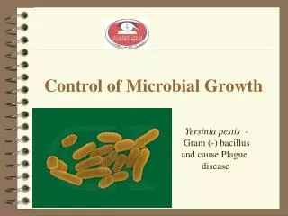
Control of Microbial Growth Tim Ho University of Alberta, Canada * The materials are mostly based on Dr. Brian Lanoil’s Microb 265 Part II tim3@ualberta.ca
Objectives • Physical agents • Mechanical removal methods • Chemical agents tim3@ualberta.ca Know 3 methods of microbial control Know the strategies on how drugs control the growth of microorganisms. Understand how do bacteria become resistant to antibiotics.
Characteristics of antimicrobial drugs: • With minimal side effects • Therapeutic dose: the amount of drug required for treatment or the desired effect. • Broad spectrum activity: against a wide variety of pathogens or do not know the specific bacteria that want to target. • Chemotherapeutic agents can be synthetic or semi-synthetic. tim3@ualberta.ca
Dilution Susceptibility test • Each test tube containing different concentrations of drug - MIC: minimum inhibitory concentration - MLC: minimum lethal concentration Low [drug] high [drug] tim3@ualberta.ca + + + + + + + - - - 4
Disk diffusion test • Kirby-Bauer method • Drug diffuses from disk into agar, establishing concentration gradient • Measure the diameter of clear zone (no growth) around disks -> determine MIC and MLC tim3@ualberta.ca Large clear zone = sensitive No or small clear zone = resistant 5 Image:http://www.biotopics.co.uk/microbes/penici.html
Disk diffusion test tim3@ualberta.ca Image:http://www.biotopics.co.uk/microbes/penici.html
How different types of antibiotics affect cell functions tim3@ualberta.ca 7
Folic acid synthesis inhibitors tim3@ualberta.ca 8
Folic acid synthesis inhibitors • Sulfanilamide: • Competitive inhibitor of PABA • [PABA] ↑= rate of folic acid biosynthesis ↓ Image: Fdardel, 2011 tim3@ualberta.ca 9
DNA gyrase inhibitors tim3@ualberta.ca 10
DNA gyrase inhibitors DNA gyrase: the enzyme that introduces negative supercoils into DNA Ciprofloxacin Image: Drug Reference - Encyclopedia • Quinolones: • inhibit bacterial DNA gyrase • effective against G- urinary tract infections and respiratory infections • (eg. Bacillus anthracis) • [PABA] ↑= rate of folic acid biosynthesis ↓ tim3@ualberta.ca 11
RNA synthesis inhibitors tim3@ualberta.ca 12
RNA synthesis inhibitors • Rifamycin/ Rifampin: • block transcription by binding RNA polymerase • Not selectively toxic • → Prokaryotes and eukaryotes synthesize nucleic acids in pretty much the same way • [PABA] ↑= rate of folic acid biosynthesis ↓ tim3@ualberta.ca 13
Cell wall synthesis inhibitors • Ampicillin is protected from lactamases by co-treatment with clavulanic acid • WHY: ß-lactamases have higher binding affinity for clavulanic acid than ampicillin tim3@ualberta.ca 14 Image: Dengler et al.BMC Microbiology 2011 11:16 doi:10.1186/1471-2180-11-16
Cell wall synthesis inhibitors G + bacteria are more susceptible to ß-lactam antibiotics!! Activity is blocked by binding to transpeptidases (transpeptidase) tim3@ualberta.ca It breaks ß-lactam rings: antibiotic resistance - G+ cells: ß-lactamases are located on outside surface G- cells: ß-lactamases are in periplasmic space 15 Image: Insilico Genomics Lab Technologies. http://insilicogenomics.in/penicillin.asp
Cell wall synthesis inhibitors blocks dephosphorylation of bactoprenol phosphate blocks transpeptidization tim3@ualberta.ca blocks D-Ala peptidization 16 Image: Dengler et al.BMC Microbiology 2011 11:16 doi:10.1186/1471-2180-11-16
Protein synthesis inhibitors tim3@ualberta.ca 17
Protein synthesis inhibitors • Macrolides: • Binding to the large subunit ribosome • Tetracyclines: • - First broad-spectrum antibiotics • Blocking tRNA attachment to ribosome • effective against G- and G+ cells tim3@ualberta.ca • Aminoglycosides: • Binding to the small subunit ribosome • effective against G- cells 18
Cytoplasmic membrane inhibitors tim3@ualberta.ca 19
Cytoplasmic membrane inhibitors • Daptomycin: • Cyclic lipopeptide • Makes pore on cytoplasmic membrane • Resistance from changes in cell membrane structure • Primarily targets G+ cells (G- cells have extra outer membrane: protection) tim3@ualberta.ca 20
Anti-fungal Drugs • Fungal infections are difficult to treat • - host and pathogen have biological similarity • → drug can harm host at the same time • Target against chitin (fungal cell wall) mostly • - animals (host) don’t have chitin • Nystatin: first discovered antifungal antibiotic in 1949 by Hazen and Brown • Superficial mycoses • - Infections of outer layers of skin • - Treatment (drugs): Miconazole, Nystatin, and Griseofulvin • - Minimizes toxic systemic side effects (e.g. liver damage) tim3@ualberta.ca 21
AntiviralDrugs • Many drugs are still in development stage • Mainly target against either RNA or DNA synthesis of viral pathogen • - Structural analogs of purine or pyrimidine bases • - difficulties: viruses use metabolic machinery of the host • Protease inhibitors: against virus-specific enzymes • Interferons: stimulate production of host anti-viral proteins tim3@ualberta.ca 22