Download
1 / 118
1.21k likes | 1.25k Views
PART 2. Muscles of the Body. Superficial Muscles of the Body – Anterior View. Figure 11.7a. Superficial Muscles of the Body – Posterior View. Figure 11.7b. Muscles of the Head – Facial Expression. Muscles of facial expression Lie in the face and scalp Thin and variable in shape

E N D
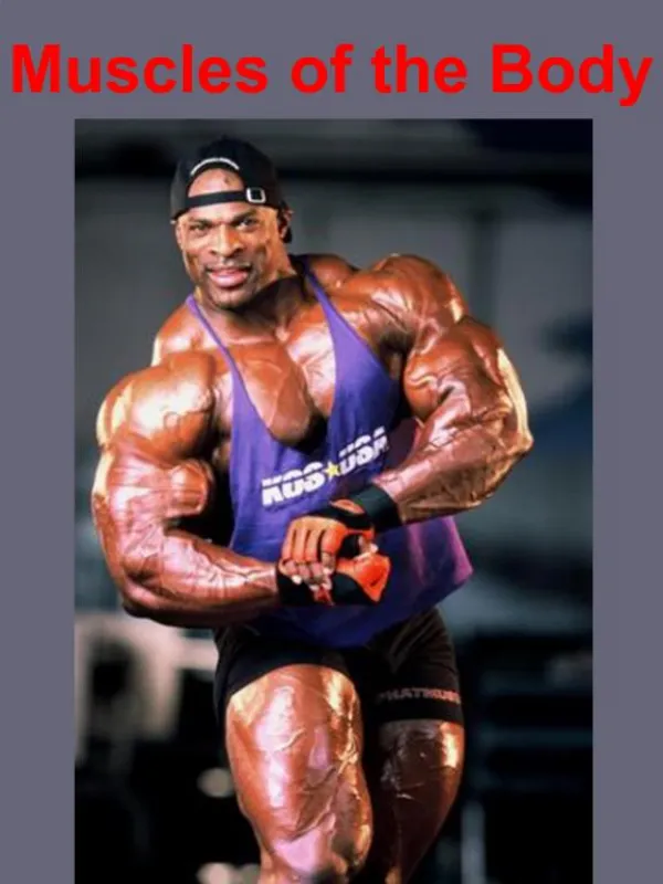
PART 2 Muscles of the Body
Superficial Muscles of the Body – Anterior View Figure 11.7a
Superficial Muscles of the Body – Posterior View Figure 11.7b
Muscles of the Head – Facial Expression • Muscles of facial expression • Lie in the face and scalp • Thin and variable in shape • Often insert in the skin – not on bones • Innervated by cranial nerve VII – the facial nerve
Muscles of the Head – Facial Expression Figure 11.8
Muscles Mastication and Tongue Movement • Four main pairs of muscles involved in mastication • Innervated by mandibular division of the trigeminal nerve (cranial nerve V) • Prime movers of jaw closure • Masseter and temporalis • Side-to-side movement • Pterygoid muscles • Compression of cheeks • Buccinator muscles
Muscles of Mastication and Tongue Movement Figure 11.9a, b
Muscles Mastication and Tongue Movement • Extrinsic muscles of the tongue • Move tongue • Laterally • Anteriorly • Posteriorly • All innervated by cranial nerve XII – the hypoglossal nerve
Muscles of Mastication and Tongue Movement Figure 11.9c
Muscles of the Anterior Neck and Throat – Swallowing • The neck is divided into anterior and posterior triangles • Anterior triangle • Divided into suprahyoid and infrahyoid muscles • Participate in swallowing • Pharyngeal constrictors • Squeeze food into the esophagus
Muscles of the Anterior Neck and Throat – Swallowing Figure 11.10a
Muscles of the Anterior Neck and Throat – Swallowing Figure 11.10b
Muscles of the Neck and Vertebral Column • Head movement • Sternocleidomastoid • Splenius capitis and splenius cervicis Figure 11.11a
Muscles of the Neck and Vertebral Column Figure 11.11b
Muscles of the Neck and Vertebral Column • Trunk extension • Deep muscles of the back • Maintain normal curvatures of the spine • Form a column from sacrum to the skull • Erector spinae group • Largest of the deep back muscles
Muscles of the Neck and Vertebral Column Figure 11.11d
Deep Muscles of the Thorax – Breathing • Deep muscles provide movements for breathing • External intercostal muscles • Lift the ribcage • Internal intercostal muscles • Aid expiration during heavy breathing
Deep Muscles of the Thorax – Breathing • Diaphragm • Most important muscle of respiration • Flattens as it contracts • Increases the volume of the thoracic cavity
Deep Muscles of the Thorax – Breathing Figure 11.12a
Deep Muscles of the Thorax – Breathing Figure 11.12b
Muscles of the Abdominal Wall • Lateral and anterior abdominal wall • Formed from three flat muscle sheets • External oblique • Internal oblique • Transversus abdominis • Fourth muscle pair • Rectus abdominis • Inserts at the linea alba
Muscles of the Abdominal Wall Figure 11.13a
Muscles of the Abdominal Wall Figure 11.13b
Muscles of the Pelvic Floor • Pelvic floor (pelvic diaphragm) • Sheet of two muscles • Both support pelvic organs • Levator ani • Formed from iliococcygeus, puborectalis, and pubococcygeus • Coccygeus
Muscles of the Pelvic Floor Figure 11.14a
Muscles of the Perineum • Inferior to the muscles of the pelvic floor • Urogenital diaphragm formed from • Sphincter urethrae and the deep transverse perineus Figure 11.14b
Muscles of the Perineum • Muscles of the superficial perineal space • Ischiocavernosus • Bulbospongiosus • Superficial transverse perineus
Muscles of the Perineum Figure 11.14c
Superficial Muscles of the Anterior Thorax • Movements of the scapula • Pectoralis major • Pectoralis minor • Serratus anterior • Subclavius
Superficial Muscles of the Anterior Thorax Figure 11.15a
Superficial Muscles of the Posterior Thorax • Movements of the scapula • Trapezius • Levator scapulae • Rhomboid major • Rhomboid minor
Superficial Muscles of the Posterior Thorax PLAY Rotator cuff muscles: an overview (a) PLAY Rotator cuff muscles: an overview (b) Figure 11.15b
Muscles Crossing the Shoulder Joint • Movements of the arm • Deltoid • Pectoralis major Figure 11.16a
Muscles Crossing the Shoulder Joint • Movements of the arm • Latissimus dorsi • Supraspinatus • Infraspinatus • Teres minor • Teres major • Coracobrachialis • Subscapularis Movement at the glenohumeral joint: an overview PLAY Figure 11.16b
Muscles Crossing the Elbow Joint Figure 11.16a
Muscles Crossing the Elbow Joint • Posterior muscles – extensors of the forearm • Triceps brachii • Anconeus • Anterior muscles – flexors of the forearm • Biceps brachii – also supinates the forearm • Brachialis • Brachioradialis
Muscles of the Forearm • Movements of the wrist, hand, and fingers • Tendons are anchored by • Flexor and extensor retinacula • Most forearm muscles arise from the distal humerus • Movements at the wrist include • Flexion, extension, abduction, and adduction • Wrist and fingers are “operated” by muscles in the forearm
Muscles of the Forearm • Flexors • Anterior flexor compartment • Innervated by median and ulnar nerves • Originate from a common tendon • Medial epicondyle of the humerus
Superficial Anterior Muscles of the Forearm • Pronator teres • Flexor carpi radialis • Palmaris longus • Flexor carpi ulnaris • Flexor digitorum superficialis Figure 11.17a
Deep Anterior Muscles of the Forearm • Flexor pollicis longus • Flexor digitorum profundus • Pronator quadratus
Deep Anterior Muscles of the Forearm Figure 11.17b, c
Muscles of the Forearm • Extensors • Posterior compartment of the forearm • Innervated by the radial nerve • Originate at a common tendon • Lateral epicondyle of the humerus
Superficial Posterior Muscles of the Forearm • Brachioradialis – flexes forearm • Extensor carpi radialis longus • Extensor carpi radialis brevis • Extensor digitorum • Extensor carpi ulnaris Figure 11.18a
Deep Posterior Muscles of the Forearm • Supinator • Abductor pollicis longus • Extensor pollicis brevis and longus • Extensor indicus Figure 11.18b
Intrinsic Muscles of the Hand • Fine movement of the fingers • All located in the palm • Control precise movements • Include muscles of • Adduction, abduction, and opposition
Intrinsic Muscles of the Hand • Thenar muscles – ball of thumb • Abductor pollicis brevis • Flexor pollicis brevis • Opponens pollicis • Adductor pollicis • Hypothenar muscles • Abductor digiti minimi • Flexor digiti minimi • Opponens pollicis PLAY Muscles that act on the wrist and fingers: an overview
Intrinsic Muscles of the Hand Figure 11.19a
Intrinsic Muscles of the Hand • Midpalmar muscles • Lumbricals • Palmar interossei • Dorsal interossei Figure 11.19b
Intrinsic Muscles of the Hand Figure 11.19c
Muscles Crossing the Hip and Knee Joints • Thigh and leg movements • Anterior muscles • Flex the thigh and extend the leg at the knee • Posterior muscles • Extend the thigh and flex the leg