Download
1 / 44
440 likes | 555 Views
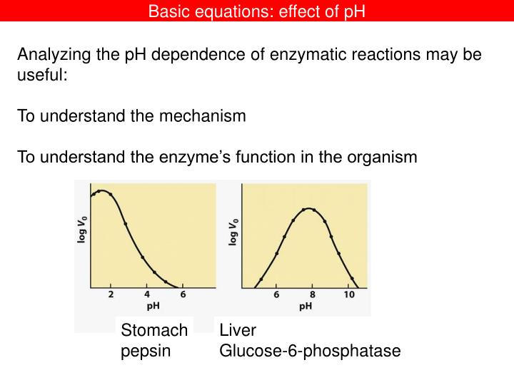
Stomach pepsin. Liver Glucose-6-phosphatase. Basic equations: effect of pH. Analyzing the pH dependence of enzymatic reactions may be useful: To understand the mechanism To understand the enzyme’s function in the organism. The questions:.

E N D
Stomach pepsin Liver Glucose-6-phosphatase Basic equations: effect of pH Analyzing the pH dependence of enzymatic reactions may be useful: To understand the mechanism To understand the enzyme’s function in the organism
The questions: 1. Which equation should we use to fit the experimental data? Basic equations: effect of pH The protonated enzyme is fully active We call it E 2. What repreents the « pKa »? The de-protonated enzyme is inactive We call it EH
pH E + S ES No reaction EH + S EHS E + products H+ H+ Ka2 Ka1 k1 k2 k-1 How to derive the Michaelis equation? Usual hypothesis initial rates mass conservation (with [S] >>[E], so [S]total = [S]free) [E]total = sum of all forms of E Steady-state [ES]=constant NOTE THAT pH=-log of FREE |H+]
pH E + S ES No reaction EH+ + S EHS+ E + products H+ H+ Ka2 Ka1 k1 k2 k-1 [E]total = sum of all forms of E [E]total = [E] + [EH+] + [ES] + [ESH+] Ka1 [H+] [E] Ka1= ------------ [E] = [EH+] ------------ [EH+] [H+] Ka2 [H+] [ES] [ES] = [ESH+] ------ Ka2= ------------ [H+] [ESH+]
pH [E]total = [E] + [EH+] + [ES] + [ESH+] Ka1 [H+] [E] Ka1= ------------ [E] = [EH+] ------------ [EH+] [H+] Ka2 [H+] [ES] [ES] = [ESH+] ------ Ka2= ------------ [H+] [ESH+] Ka2 Ka1 [E]total = [EH+] -------- + [EH+] + [ESH+] ------ + [ESH+] [H+] [H+] Ka1 Ka2 [E]total = [EH+] (-------- + 1)+ [ESH+] (------ + 1) [H+] [H+]
pH E + S ES No reaction EH + S EHS E + products H+ H+ Ka2 Ka1 k1 k2 k-1 Ka1 Ka2 [E]total = [EH+] (1+ -------)+ [ESH+] (1+ ------) [H+] [H+] X2 X1 [E]total = [EH+] X1 + [ESH+] X2 Next step: steady-state k1 [EH] [S] = (k –1 + k 2) [EHS] [EH] = (k –1 + k 2)/ k1 [EHS]/[S]
pH [E]total = [EH+] X1 + [ESH+] X2 [EH] = (k –1 + k 2)/ k1 [EHS]/[S] (k –1 + k 2)/ k1 = Km [E]total = [ESH+] Km/[S] X1 + [ESH+] X2 V = k2 [EHS] k2 [E]total V = --------------------------- Km/[S] X1 + X2 Vmax [S]/X2 V = --------------------------- Km X1/X2 + /[S]
pH Vmax [S]/X2 V = --------------------------- Km X1/X2 + /[S] Vmaxapp[S] V = ----------------- Kmapp + /[S] 1 Vmaxapp = Vmax ---------------- Ka2 1 + ------ [H+] Ka1 1 + ------ Vmaxapp 1 Vmax [H+] ----- = ------- ------------ Kmapp = Km----------------- Ka1 Kmapp 1 + ------ Km Ka2 [H+] 1 + ------ [H+] Vmaxapp ------------ is a function of the Ka of the FREE enzyme (it can be measured directly) Kmapp
pH 1 Vmaxapp = Vmax ---------------- Ka2 1 + ------ [H+] Ka1 Vmaxapp 1 Vmax 1 + ------ ----- = ------- ------------ [H+] Ka1 Kmapp = Km----------------- 1 + ------ Kmapp Km Ka2 [H+] 1 + ------ [H+] Last step: verify that the equation describes our experiment: In acid medium [H+]>>Ka2 Vmaxapp = Vmax In alkaline medium,[H+]<<Ka2 Vmaxapp = 0 OK ! ALLWAYS VERIFY THAT YOUR FINAL EQUATION IS CORRECT
pH In this reaction scheme, H+ is an mixed-type activator (because it binds both to the free enzyme and to the ES comples) Practical hint: the « kinetic » pKa may be obtained from the log-log plot:
pH What happens if the enzyme is inactive while protonated? H+ is an mixed-type inhibitor. You may use directly the equation for mixed inhibition.
What happens if the enzyme is active at alkaline, but not at acidic pH?
Suppose that the enzyme is not protonated/deprotonated, but the substrate is. TDP X = OH AZT-DP X = N3 3’-NH2-TDP X = NH2 3’-NH3+-TDP is not a substrate
Basic equations: effect of pHA MORE COMPLICATED SITUATION pKa1 pKa2
Basic equations: effect of pHA MORE COMPLICATED SITUATION • The reversible effect of pH on an enzyme can be described in a scheme that is only slightly more complex than a classic inhibition scheme. Ionizations “inhibit” either the free enzyme or the enzyme-substrate complex. k1 k-1 ionizations in the enzyme-substrate complex ionizations in the free enzyme or free substrate
[H+] KES2 ------ + 1 + ------ KES1 [H+] Vmax [H+] KE2 Vmaxapp = ---------------- ------ + 1 + ------ KE1 [H+] Kmapp = Km-------------------- [H+] KES2 ------ + 1 + ------ KES1 [H+] Vmaxapp 1 Vmax ----- = ------- --------------- [H+] KE2 Kmapp Km ------ + 1 + ------ KE1 [H+]
Acid-base properties of amino acids side chains • Attention! • Strong acids (HCl) and strong bases (NaOH) do not have pKa • (they are totally dissociated) • H+ in solution is H3O+ Same pKa scale for acids and bases AH A- + H+ -COOH COO- + H+ BH+ B + H+ -NH3+ NH2 + H+ Acide/base Eq de HENDERSON-HASSELBALCH pH = pKa + log10 [ A- ]/[ AH ]
Acid-base properties of amino acids side chains Eq de Henderson-Hasselbalch pH = pKa + log10 [ A- ]/[ AH ] [AH] Binding curve [AH] Titration curve
Acid-base properties of amino acids side chains tautomers Histidine is aromatic both protonated and unprotonated Histidine pKa 6.5 Lysine pKa 9.5 Arginine pKa 12
Acid-base properties of amino acids side chains Aspartate et Glutamate pKa 4.4 COOH terminal pKa 3.0 Cystéine pKa 8.5 Tyrosine pKa 10 NH2terminal pKa 8.0
Acid-base properties of amino acids side chains 0 +1 The slope is the same irrespective of the pKa, UNLESS there is a cooperative proton binding
Acid-base properties of amino acids side chains Macrocryptates The facilitation of the second protonation of 10 represents a positive cooperativity, in which the first proton and the effector molecule water set the stage both structurally and energetically for the fixation of a second proton. SUPRAMOLECULAR CHEMISTRY - SCOPE AND PERSPECTIVES MOLECULES - SUPERMOLECULES - MOLECULAR DEVICES Nobel lecture, December 8, 1987 by JEAN-MARIE LEHN
Acid-base properties of amino acids side chains + + + + + + - - - - - - • L’environnement apolaire des chaînes latérales favorise la forme non-ionisée • pKa augmente pour les COOH et diminue pour les NH2 • 2. La proximité d’une chaîne latérale chargée modifie le pKa His dans une protéine
Acid-base properties of amino acids side chains NH2-CH2-COOH Glycine pKa are 2.35 and 9.78 The dipeptide glycylglycine pKa are 3.12 and 8.17 NH3+-CH2-COO- NH2-CH2-COO- NH3+-CH2-COOH NH3+-CH2-CO-NH-CH2 -COOH NH3+-CH2-CO-NH-CH2 -COO- NH2-CH2-CO-NH-CH2 -COO-
Acid-base properties of amino acids side chains Glycine pKa of the –NH2 group is 9.78, while in glycinamide is 8.2 NH3+-CH2-COO- NH2-CH2-COO- pKa 9.78 NH3+-CH2-CO-NH2 NH2-CH2-CO-NH2pKa 8.2 From the pKa we may calculate the free energy associate to the salt bridge
Acid-base properties of amino acids side chains NH3+-CH2-COO- NH2-CH2-COO- pKa 9.78 NH3+-CH2-CO-NH2 NH2-CH2-CO-NH2pKa 8.2 From the pKa we may calculate the free energy associate to the salt bridge
Acid-base properties of amino acids side chains NH2-CH2-COOH Glycine pKa are 2.35 and 9.78 The dipeptide glycylglycine pKa are 3.12 and 8.17 NH3+-CH2-COOH NH3+-CH2-COO-pKa 2.35 NH3+-CH2-CO-NH-CH2 -COOH NH3+-CH2-CO-NH-CH2 -COO-pKa 3.12 The ionic interaction decreases the pKa, more for glycine since charges are closer
Acid-base properties of amino acids side chains pKa 8.17 NH3+-CH2-CO-NH-CH2 -COO- NH2-CH2-CO-NH-CH2 -COO- La glycine a des valeurs de pKa de 2.35 et 9.78 Le dipeptide glycylglycine a des valeurs de pKa de 3.12 et de 8.17 NH2-CH2-COOH NH3+-CH2-COO- NH2-CH2-COO- pKa 9.78 The loss of the stabilizing interaction is more important for glycine since charges are closer
How to measure side chains pKa in proteins? Why are pKs important? Protein structure, stability and solubility depends on the charge on ionizable residues Catalytic residues at enzyme active sites are 65% charged, 27% polar and 8% nonpolar Catalytic residues at enzyme active sites are 18% His, 15% Asp, 11% Arg, and 11% Glu (Barlett, Porter, Borkakoti, & Thornton, JMB, 324, 105 (2002))
1. Propriétés acido-basiques pK values for the ionizable residues in folded RNase Sa Residue (N & T)Measured pK C-term(3.8)2.42 Asp 01 (4.0)3.44 Asp 173.72 Asp 254.87 Asp 332.39 Asp 797.37 Asp 843.01 Asp 933.09 Glul4(4.4)5.02 Glu414.14 Glu543.42 Glu743.53 Glu783.17 His 53 (6.3)8.27 His 856.24 N-term (7.5)9.14 Tyr 30 (9.6)11.3 Tyr 4910.6 Tyr 51, 52, 55> 11.5 80, 81, 86
Highly perturbed pKa values in enzymes • COOH In a low polarity environment • ionization is disfavoured pKa • COO– - - - - +H3N • ionization is stabilized via salt bridge pKa of carboxyl function pKa of amino function • NH2 In a low polarity environment • charged group is disfavoured pKa Examples of perturbed pKa values: lysozyme Glu-35 (pKa = 6.5) acetoacetate decarboxylase Lys-NH3+ (pKa = 5.9) papain His-159 (pKa 3.4) the environment surrounding an ionizable group can greatly influence its ionization
How to measure side chains pKa in proteins? • It is of course more easy to calculate, but we want te MEASURE! • The proteins should be native (which exclude experiments <4 or >10 • Most easy end interesting is the histidine pKa • How? • General method: NMR (but the protein should be small <200 aa) • Specific methods • Example: HisH+ decreases the fluorescence intensity of a neighbouring Tryptophane
How to measure side chains pKa in proteins? C-H stable C-H stable 13C Signals typical for protéine native N-H rapidly exchange in D2O and therefore the signal is lost 1H-RMN Aromatic protons Histidines are identified by site-directed mutagenesis
How to measure side chains pKa in proteins? Example 1, phospholipase C of Bacilus cereus Phospholipase C This bond is hydrolyzed (nucleophilic atak on phosphorous Mécanisme similaire à celui e la RNaseA
Comment MESURER le pKa des chaînes latérales dans les protéines? ex2 Histidine Position H32 7.6 Site actif H82 6.9 Site actif H61 — Enfuie H81 _ Enfuie H92 5.4 Surface de la protéine H227 6.9 Surface de la protéine Tible 2. The pK, values of the histidines of B. cereus Pl-PLC pKa dans des peptides: 6.5
How to measure side chains pKa in proteins? Example 1, phospholipase C of Bacilus cereus Determination of pKa values of the histidine side chains of phosphatidylinositol-specific phospholipase C from Bacillus cereus by NMR spectroscopy and site-directed mutagenesis. Protein Sci. 1997 Sep;6(9):1937-44. Liu T, Ryan M, Dahlquist FW, Griffith OH. Institute of Molecular Biology, University of Oregon, Eugene 97403, USA. Two active site histidine residues have been implicated in the catalysis of phosphatidylinositol-specific phospholipase C (PI-PLC). In this report, we present the first study of the pKa values of histidines of a PI-PLC. All six histidines of Bacillus cereus PI-PLC were studied by 2D NMR spectroscopy and site-directed mutagenesis. The protein was selectively labeled with 13C epsilon 1-histidine. A series of 1H-13C HSQC NMR spectra were acquired over a pH range of 4.0-9.0. Five of the six histidines have been individually substituted with alanine to aid the resonance assignments in the NMR spectra. Overall, the remaining histidines in the mutants show little chemical shift changes in the 1H-13C HSQC spectra, indicating that the alanine substitution has no effect on the tertiary structure of the protein. H32A and H82A mutants are inactive enzymes, while H92A and H61A are fully active, and H81A retains about 15% of the wild-type activity. The active site histidines, His32 and His82, display pKa values of 7.6 and 6.9, respectively. His92 and His227 exhibit pKa values of 5.4 and 6.9. His61 and His81 do not titrate over the pH range studied. These values are consistent with the crystal structure data, which shows that His92 and His227 are on the surface of the protein, whereas His61 and His81 are buried. The pKa value of 6.9 corroborates the hypothesis of His82 acting as a general acid in the catalysis. His32 is essential to enzyme activity, but its putative role as the general base is in question due to its relatively high pKa.
How to measure side chains pKa in proteins? Example 2, the Ribonuclease A The pH titration curves of the six histidines in B. cereus PI-PLC. The pKa values were determined by non-linear regression fitting, using the Henderson-Hasselbalch equation
How to measure side chains pKa in proteins? Example 3, the perturbation by charged ressidues Long-range surface charge-charge interactions in proteins. Comparison of experimental results with calculations from a theoretical method. J Mol Biol. 1993, 232, 574-83. Loewenthal R, Sancho J, Reinikainen T, Fersht AR. MRC Unit for Protein Function and Design, Cambridge, England. PDB 1SBT 24.5 A His64 Asp36 Lys136
How to measure side chains pKa in proteins? Example 3, the perturbation by charged ressidues

![Solving [Specific Classes of] Linear Equations using Random Walks](https://cdn0.slideserve.com/33886/solving-specific-classes-of-linear-equations-using-random-walks-dt.jpg)