Download
1 / 66
720 likes | 2.18k Views
Prosthetic valves. Types Selection Complications . Types . Bioprosthetic valves Heterograft ( xenograft ) Bovine porcine Homograft (allograft) Autograft Pericardial Pulmonary (Ross) Mechanical Caged ball valve Tilting disc valve Bileaflet valve.

E N D
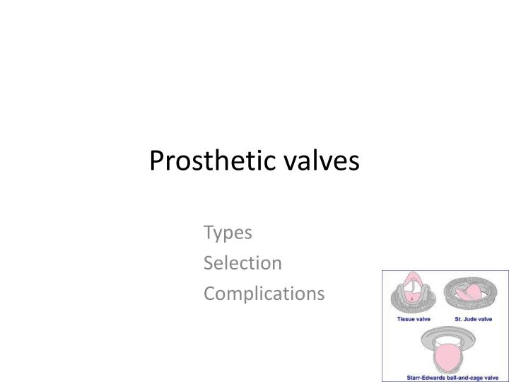
Prosthetic valves Types Selection Complications
Types • Bioprosthetic valves • Heterograft (xenograft) • Bovine • porcine • Homograft (allograft) • Autograft • Pericardial • Pulmonary (Ross) • Mechanical • Caged ball valve • Tilting disc valve • Bileaflet valve
Heterografts(xenografts) The Hancock M.O. II aortic bioprosthesis (porcine) • Stentless • Porcine • Toronto SPV valve, medtronicfreestyle valve • Stented - facilitate implant, maintain 3D relationship,more physiological flow • Porcine • Hancock , carpenteiredwards, medtronic • Bovine • Stented bovine p prosthesis Carpentier-Edwards Duralex mitral bioprosthesis
Advantages - No need of anticoagulation after 1st 3 m Little hemolysis Disadvantages Limited ,uncertain durability Cuspal tear Perforation degeneration Rapid deterioration esp children Fibrin depostn Ca++ 10-30% need re op in 10 yr 30-60% need re op in 15 yr Small size have poor hemodynamics Tissue heterograft
Bioprosthetic valve Preferred in • Pregnancy • Bleeding Diathesis • Age> 70 years • Poor compliance
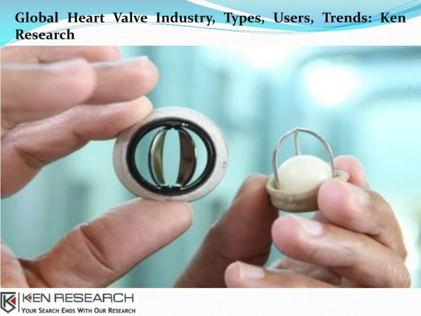
Mechanical valves • Caged ball valve Advantages • Oldest • durabiltyupto 40 yr Disadvantages • high profile • hemolysis • high thrombogenecity • Poor hemodynamics in small sizes Unique features • Occluder travels completely out of the orifice, reduces thrombus & pannus growing from the sewing ring • Continuously changing points of contact of the ball reduces the wear & tear in any one area • Thrombogenic risk 4-6% / year
Starr Edwards Valve • not suitable - for the mitral position in patients with a small left ventricular cavity - for the aortic position in those with a small aortic annulus - those requiring a valve-aortic arch composite graft
Tilting disc valve- monoleaflet • - Medtronic Hall valve - Omnicarbon (Medical CV) - Monostrut (Alliance Medical Technologies) - Bjork-Shiley valves • Adv • low profile • Good hemodynamics even in small sizes • Excellent durability • Permit central laminar flow • Medtronic hall valve • Titanium housing • teflon sewing ring • carbon coated disc • disadv– • Anticoagulation mandatory • higher risk of thrombosis than cage ball v • sudden catastrophic valve thrombosis
St. Jude Medical mechanical heart valve Bileafletvalve Adv – • Low bulk - flat profile • Less thrombogenicy • Central laminar flow • two semicircular discs that pivot between open and closed positions • No need for supporting struts • Good hemodynamics even in small sizes • 2 lat ,1 central minor orifice , no chance of sudden catastro thrombosis Disadv- • Anticoagulation mandatory • risk of thrombosis • Carbomedics • Titanium housing • Pyrolytic carbon
TTK chitra • tilting disc valve - metallic housing (cobalt based wrought alloy) - circular disc high molecular weight polyethylene - A polyester suture ring • Hemodynamically comparable to other mechanical valves • valve related complications are similar
Desired valves • Mechanical valves - preferred in young patients who have a life expectancy of more than 10 to 15 years who require long-term anticoagulant therapy for other reasons (e.g., atrial fibrillation) • Bioprostheticvalves preferred in patients who are elderly have a life expectancy of less than 10 to 15 years who cannot take long-term anticoagulant therapy • A bileaflet-tilting-disk or homograft prosthesis is most suitable for a patient with a small valvular annulus in whom a prosthesis with the largest possible effective orifice area is desired.
Radiologic Identification Starr-Edwards caged ball valve Radiopaque base ring Radiopaque cage Three struts for the aortic valve; 4 struts for the mitral or tricuspid valve Silastic ball impregnated with barium that is mildly radiopaque (but not in all models)
TTE – stenosis Valve area calculations • Continuity equation Area Ao prosthesis = (diameter sewing ring)² x 0.785xLVOT VTI/ Ao prosthesis VTI Area mitral prosthesis= (diameter LVOT)²x 0.785xLVOT VTI/ VTI mitral prosthesis • Pressure Half time ( mitral valve prosthesis) • Dimensionless index- LVOT velocity/ aortic prosthesis velocity < 0.23 indicates prosthetic valve stenosis
Prosthetic Valve regugitation • Mitral – velocity2.5m/sec - jet area 2cm² • Aortic -- aortic PHT≤ 250m/sec -- flow reversal in aorta
Normal Doppler Values of Prosthetic Valves Aortic Position Mitral Position Velocity MeanGr Starr Edward 1.8±0.5 7±2 St Jude 1.5±0.3 5±2 Medtronic Hall 1.6±0.3 5±2 Aortic Homograft 1.5±0.4 4±2 Hancock 1.5±0.3 5±2 Carpentier’s 1.5±0.3 5±2 Velocity Mean Gr Starr Edward 3.1±0.5 24±4 St Jude 3.0±0.8 11±6 Medtronic Hall 2.6±0.3 12±3 Aortic Homograft 0.8±0.4 7±3 Hancock 2.4±0.4 11±2 Carpentier’s 2.4±0.5 14±6
Importance of TEE • higher-resolution image than TTE • size of vegetation defined more precisely • peri annular complications indicating a locally uncontrolled infection (abscesses, dehiscence, fistulas) detected earlier • limitation -inability to detect aortic prosthetic-valve obstruction or regurgitation, especially when a mitral prosthesis is present
Cinefluoroscopy • Structural integrity • Motion of the disc or poppet • excessive tilt ("rocking") of the base ring - partial dehiscence of the valve • Aortic valve prosthesis - RAO caudal - LAO cranial Mitral -- RAO cranial
Fluoroscopy of a normally functioning CarboMedicsbileaflet prosthesis in mitral position A=opening angle B=closing angle
St. Jude medical bileaflet valve • Mildly radiopaque leaflets are best seen when viewed on end • Seen as radiopaque lines when the leaflets are fully open • Base ring is not visualized on most models
MRI • Not useful in assessing prosthetic-valve structure • used only when prosthetic-valve regurgitation or paravalvular leakage is suspected but not adequately visualized by echocardiography
Cardiac Catheterization • measure the transvalvular pressure gradient, from which the EOA can be calculated • can visualize and quantify valvular or paravalvularregurgitation
patient-prosthesis mismatch • When the effective prosthetic valve area, after insertion into the patient less than that of a normal valve (Rahimtoola in 1978) • EOA indexed to BSA is less than 0.85 cm2/m2 • EOA (echo) differs from geometric orifice area (measured directly) • EOA for each prostheses type & size obtained in literature from pts normally functioning prostheses • Average if > 1 value -- mild (0.9 - 1 cm² /m² -- moderate (0.6 - 0.9 cm2/m² -- severe (iEOA < 0.6cm²/m² (Rahimtoola)
in-vitro area of the majority of valve prostheses ( int diameter <23 mm) < that of the normal human valve area • the in-vivo prosthetic area further reduced by IVS hypertrophy, progressive endothelialization and tissue ingrowth (Aortic prosthetic devices may be functionally stenotic)
three-step algorithm • Step 1: Calculation of the patientBSA. Step 2: Reference to the specific table for identification of the adequate valvular EOA according to the patientBSA. Step 3: Selection of the most appropriate type and size of valve prosthesis according to the target iEOA
Valve Thrombosis Incidence of 0.1 to 5.7 % per patient-year <0.2% per year for mech valves <0.1% bioprosthetic valves • small thrombus, at the hinge portion of a bileaflet valve obstruct the mechanism • tilting disk -- a much larger thrombus to prevent function • Ball and cage valves – less susceptible occluder has no contact at all with the valve housing for a portion of every cycle Clinical • Non obstructive- incidental/embolic phenomenon • Partial obstruction- dyspnea,systemic embolism , fever • Severe obstruction- overt heart failure
Fibrinolytic therapy - Rt sided thrombosis 80-100% success rate • Surgery for fibrinolysis failure/symptoms > 3 wk • Surgery – Lt sided thrombosis, large clot burden
FIBRINOLYTIC PROTOCOLheart 2007;93:137-142 • 2 types of protocol -rescue fibrinolysis (short protocol for unstable pt) - long protocol for stable pt • Short protocol - r tPA 10 mg bolus + 90 mg in 90 min or - SK 15lac in 60 min • Long protocol -- SK- 5lac u in 20 min f/b 15lac u for 10 hr -- rtPA -- 10 mgbolus f/b 90mg/hr for 9 hrs • Urokinase • High dose: 4,500 IU/kg/h for 12 h without heparin • Low dose: 2,000 IU/kg/h with heparin for 24 h
Embolisation • cerebral embolization CT normal/infarctwarf & heparin – 72 hrs APTT lower therapeutic level till the desired INR • anticoagulantion delayed for at least 7 to 14 days - ICH, extensive cerebral infarction OAC
If embolic event occurs while the patient is on adequate antithrombotic therapy • If on warfarin with INR of 2.0 to 3.0: increase dose to achieve INR of 2.5 to 3.5 • If on warfarin with INR of 2.5 to 3.5: add aspirin 50 to 100 mg/d • If on warfarin with INR of 2.5 to 3.5, plus aspirin 80 to 100 mg/d: aspirin dose may also need to be increased to 325 mg/d • If on aspirin 325 mg/d: switch to warfarin with goal INR of 2.0 to 3.0
Excessive Anticoagulation • vit K 2.5 mg daily until the INR is acceptable • fresh frozen plasma • Human recombinant factor VIIa, 15 to 19 g/kg (INR >10.0 with bleeding)
Structural Failure of Bioprosthetic Valves • About 30 % of heterograft bioprosthetic valves and 10 to 20 % of homograft valves require replacement within 10 to 15 years because of structural failure • severe regurgitation due to a tear or rupture of one or more of the valve cusps • calcified and rigid valves • Rarely severe valvularstenosis
Structural deterioration • Higher incidence patients <40 years & with mitral prostheses • gradual onset of dyspnea and other symptoms of heart failure • Bioprosthetic-valve regurgitation or stenosis can be detected by auscultation • valve dysfunction assessed by echocardiography or catheterization
Hemolysis • Incidence - 6% • Subclinical intravascular hemolysis • severe hemolytic anemia uncommon & suggests paravalvular leakage due to partial dehiscence of the valve or infection • Patients with a caged-ball valve / multiple prosthetic valves have an increased incidence & severity of hemolysis.
Hemolysis • Pts with hemolytic anemia treated with iron & folate supplements or blood transfusion - decreased blood viscosity & increased COP a/w anemia increase the hemolysis • Paravalvular leakage & severe hemolysis – valve replacement or repair
Para valvular leak • improper implantation of a valve • A heavily calcified annulus is a risk factor for paravalvular leaks -- incomplete debridement of calcium compromises both suture placement and valve seating • Active endocarditis is also a risk factor • Late paravalvular leaks are suggestive of prosthetic valve endocarditis • generally result in hemolysis • In the absence of a paravalvular leak, a normally functioning modern valve should not result in hemolysis
Paravalvular Regurgitation • mild or moderate paravalvular leakage - asymptomatic , may have only a mild hemolytic anemia - can be observed carefully with serial echo • severe paravalvular leakage - usually have symptoms of heart failure or severe anemia - should be treated with surgical repair or replacement of the valve
PVE (2-6%) salient features • Endovascular, microbial infection occurring on parts of a valve prosthesis or on reconstructed native heart valves , with or without implantation of an annular ring • early PVE is 5% higher in surgery during active IE • Diagnostic approach, surgical indications same