Download
1 / 61
610 likes | 771 Views
Introduction to Microbiology/Prokaryotes. Dr. Cory L. Blackwell August 26, 2014. Changing Paradigms. Before the 1880’s it was believed that diseases came from demons and witchcraft

E N D
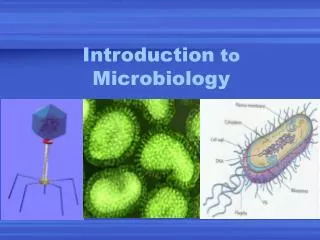
Introduction to Microbiology/Prokaryotes Dr. Cory L. Blackwell August 26, 2014
Changing Paradigms • Before the 1880’s it was believed that diseases came from demons and witchcraft • With the new concept of biogenesis and the realization that microorganisms can physically and chemically change organic materials, scientists/physicians began to develop The Germ Theory of Disease.
GERM THEORY OF DISEASE • The Germ Theory of Disease states that a particular microbe might cause a specific disease • 1865 Joseph Lister, English surgeon, began treating surgical wounds with phenol (acid) resulting in the significant reduction of infections and deaths • 1876 Robert Koch discovered that the bacterium Bacillus anthracis was present in the blood of cattle that died of the disease anthrax • Koch eventually established Koch’s Postulates
Koch’s Postulates • The same pathogen must be isolated in each case of the disease • Pathogen must be isolated from the diseased host and grown in a pure culture • The pathogen from the culture must cause the same disease when used to inoculate a healthy organism • Microorganism must be isolated from the newly diseased animal and be identical to the original pathogen
Prokaryote or Eukaryote? Prokaryotes Eukaryotes Humans Plants Yeast Fungi Amoebas • Bacteria • Archaea
Prokaryote and Eukaryote • Prokaryotes and Eukaryotes have four common features • Cell membrane • Cytoplasm • Nucleic Acid • Ribosomes • Both utilize similar chemical reactions to metabolize food, build proteins, and store energy
Eukaryotic or Prokaryotic • Prokaryotes and Eukaryotes differ in several ways:
DNA Location • DNA of Eukaryotes are found in the cell’s nucleus—membrane bound organelle that contains the genetic material • DNA of Prokaryotes is not enclosed in a membrane bound organelle
DNA Location Prokaryote Eukaryote
DNA Structure • Eukaryotic DNA is bound by chromosomal proteins called histones and is found in multiple chromosomes • Prokaryotic DNA is usually takes the form of a singular circularly arranged chromosome. No histones are present
Prokaryotic DNA Eukaryotic DNA
Organelles • Eukaryotes possess membrane-enclosed organelles • Mitochondria, endoplasmic reticulum, golgi apparatus, lysosomes, nucleus, etc. • Prokaryotes are void of an membrane-enclosed organelles
Cell Wall Composition • Prokaryotes contain peptidoglycan (complex polysaccharide) within its cell walls • Lipotechoic acids (LTA) • Lipopolysaccharides (LPS) • Eukaryotes cell wall (only in plant cells and fungi) are made of simple molecules (cellulose and chitin)
Cell Wall Composition Eukaryotic Cell Membrane Prokaryotic Cell Wall
Growth and Division • Prokaryotes divide by binary fission • Requires relatively few structure and processes • Eukaryotes divide utilizing mitosis • More complex than binary fission • Interphase, prophase, metaphase, anaphase, telophase
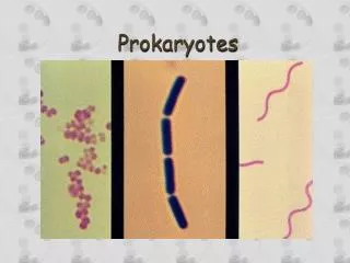
Prokaryotes • Prokaryotes are subdivided into two groups • Bacteria • Archaea • Both bacteria and archaea are unicellular organisms • Although bacteria (constitutes the majority of prokaryotes) and archaea look similar, their chemical composition is different • Archaealack peptidoglycan in their cell walls • Archaea usually live in extreme environments • Methanogens • Extreme Halophiles • Hypothermophiles
Bacterial Size, Shape, and Arrangement • Most bacteria range between 2 to 8 µm in length • That is roughly 1,000X less than the size of an ant • Bacteria have three different cellular shapes • Coccus“spherical” • Bacillus “rod-shaped” • Spiral “spiral-shaped”
Cocci • Bacterial cells that are cocci can be oval, elongated, or flat on one side
Cocci • The cocci are also distinguished based on how the bacteria group with one another (bacterial arrangement)
Spatial Arrangement of Cocci • Diplo—pairs of bacteria • Strepto—chains of bacteria • Staphyl—grapelike clusters • Tetrads—groups of four • Sarcinae—cube like structures consisting of 8 bacteria
Bacilli • Bacilli divide only across their short axis, therefore there are less arrangement groupings • Diplo—pairs of bacteria Bacillus anthracis
Bacilli • Bacilli divide only across their short axis, therefore there are less arrangement groupings • Diplo—pairs of bacteria • Strepto—chains of bacteria arranged from tip to tail
Bacilli • Coccobacilli—combination of rod and oval shapes Brucellamelitensis
Spiral • Spiral bacteria form twists and are never found in a straight conformation • Spiral bacteria come in three varieties: • Vibrio • Spirillum • Spirochete
Vibrioare in the shape of curved rods Vibrio cholera
Spirilla have a helical “corkscrew” shape • Have rigid bodies Camplyobacterjejuni
Spirochetes are helical in nature • Bodies are FLEXIBLE • Difference between spirilla and spirochetes Treponemapallidum
Bacterial Shapes • Bacterial shape is determined by heredity • If the parent cells are of a certain shape their progeny will be of similar shape • Most bacteria are monomorphic, or maintain a single shape • Although the environment may play a role in changing a bacterium’s shape • When a bacterium’s shape is altered it is referred to as pleomorphic(Corynebacterium) • Leads to difficulty in identifying bacteria
Critical Thinking • The name of a bacterium is often associated with its shape. Draw these organisms based on their names • Streptococcus pneumoniae • Staphlycoccusaureus • Bacillus anthracis • Vibrio cholera
External Cell Wall Structures • The cell wall is a crucial component of the bacteria • There are several structures that line the outside of the cell wall that have a variety of functions
Glycocalyx • Glycocalyx—substance that is secreted on the surface of the cell walls by prokaryotes • Glycocalyx (sugar coat) is a sticky, gelatinous polymer that is composed of a polysaccharide, polypeptide, or both • If the glycocalyx is organized and firmly attached to the cell wall, it is referred to as a capsule • Inversely, if the glycocalyx is unorganized and loosely attached it is called a slime layer
Glycocalyx Capsule Slime Layer
Glycocalyx • The glycocalyx serves a variety of functions • Helps prevent the phagocytosis (ingestion by immune cells) of bacterial cells, thus aiding in their pathogenicity • Forms biofilms that not only shield the bacteria from external stimuli (salt concentrations or antibiotics) but also aid in the communication between the bacteria • Helps anchor bacteria to specific surfaces • Can also be used as a source of nutrients for the bacterial cells
Flagella • Some prokaryotes have flagella which are long filamentous appendages that aid in bacterial motility (ability of a bacteria to move by itself) • H antigen—flagellar protein that aids in distinguishing serovars of bacteria • Serovars—variations within a species • The type of flagella can be categorized based on its arrangement on the bacteria
Flagella • Atrichous—bacteria that lacks flagella • Peritrichous—distributed all around the cell • Monotrichous—single flagellum at one end • Lophotrichous—several flagella at one end • Amphitrichous—flagella at both ends
Flagella Movement • The movement of the flagella propels the bacteria towards a favorable environment or away from an unfavorable environment • This movement is referred to as taxis • Chemotaxis—movement towards a chemical • Phototaxis—movement towards light
Other Bacterial Projections • Axial Filaments—bundles of filaments that arise at the end of the cell beneath the outer sheath. Spiral around the entire cell • Bacteria with axial filaments move in a corkscrew shape motion
Other Bacterial Projections • Fimbriae—small hair-like appendages that adhere to each other and surfaces. Involved in forming biofilms • Placement is either at the ends of the bacteria or around the entire bacteria
Other Bacterial Projections • Pili—appendages that are involved in motility and DNA transfer • The transfer of DNA from one bacteria to another is termed conjugation • The sex pili (F+) binds to the recipient bacteria and injects genetic material Sex Pilus (F+)
Bacterial Cell Wall • The cell wall is a complex, semi-rigid structure that surrounds the underlying, fragile plasma membrane • Major function of the cell wall is to prevent bacteria cells from rupturing due to osmotic pressure • They also help the bacteria to maintain its shape. • Point of anchor for the external appendages
Cell Wall Composition • The main component of the bacterial cell wall is a macromolecule called peptidoglycan • Peptidoglycanconsists of repeating a disaccharide attached to polypeptides to form a lattice around the cell
Cell Wall Composition • The cell wall, depending on its composition, exhibits different characteristics • Because of this, bacteria can be placed in two different groups: Gram-Positive bacteria and Gram-Negative bacteria • The major difference between Gram-Positive and Gram-Negative bacteria is the peptidoglycan composition • G(-) have an outer membrane, inner membrane, and contain periplasm • G(+) lack outer membrane and do not contain periplasmic space