Download

1 / 28
280 likes | 548 Views
Non-small-cell lung cancer. DR.Mohammed Awad. Incidence. The most common cause of cancer mortality in both sex (1/3 of cancer deaths) About 90% of lung cancer mortality among men (and 80% among women) is attributable to smoking.

E N D
Non-small-cell lung cancer DR.Mohammed Awad
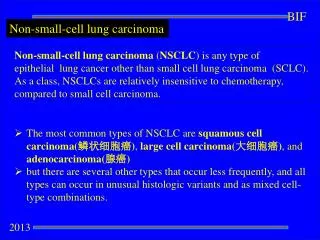
Incidence • The most common cause of cancer mortality in both sex (1/3 of cancer deaths) • About 90% of lung cancer mortality among men (and 80% among women) is attributable to smoking. • Non-small-cell lung cancer (NSCLC) accounts for 80% of all cases.
Risk Factors • Cigarette, pipe, or cigar smoking. • Exposure to second-hand smoke, radon, arsenic, asbestos, chromates, chloromethyl ethers, nickel, polycyclic aromatic hydrocarbons, radon progeny, other agents, and air pollution. • Radiation therapy to the breast or chest.
Risk Factors Cont. • For smokers, the risk for lung cancer is on average tenfold higher than in lifetime nonsmokers. • The risk increases with the quantity of cigarettes, duration of smoking, and starting age. • Smoking cessation results in a decrease in precancerous lesions and a reduction in the risk of developing lung cancer. • Former smokers continue to have an elevated risk for lung cancer for years after quitting. • Asbestos exposure may exert a synergistic effect of cigarette smoking on the lung cancer risk
Screening • In patients considered at high risk for developing lung cancer, no screening modality for early detection has been shown to alter mortality. • Studies of lung cancer screening with chest radiography and sputum cytology have failed to demonstrate that screening lowers lung cancer mortality rates.
Screening Cont. • Published studies of newer screening technologies such as low-dose computed tomography (CT) scans and biomarker screenings report primarily on lung cancer detection rates and do not present sufficient data to determine whether the newer technologies will benefit or harm people. • Currently, randomized trials are evaluating low-dose spiral CT scanning.
WHO/INTERNATIONAL ASSOCIATION FOR THE STUDY OF LUNG CANCER HISTOLOGIC CLASSIFICATION OF NSCLC
Squamous cell carcinoma. • Adenocarcinoma. • Large cell carcinoma. • Adenosquamous carcinoma. • Carcinomas with pleomorphic, sarcomatoid, or sarcomatous elements. • Carcinoid tumor. • Carcinomas of salivary-gland type. • Unclassified carcinoma
Staging and Risk Assessment • Complete history and physical examination, CT scan of the chest and upper abdomen. • MRI of the brain if abnormal neurologic findings (can be substituted by CT scan) • Bone scan in the presence of bone pain, elevated serum calcium, or elevated alkaline phosphatase levels.
Patients With Potentially Curative Treatment • Biopsy of mediastinal lymph nodes 1 cm in short axis. • Biopsy of mediastinal lymph nodes can be obtained by mediastinoscopy, transbronchial needle aspiration, ultrasound-guided bronchoscopy with fine needle aspiration, and /or endoscopic esophageal ultrasound-guided fine needle aspiration. • MRI of the brain for clinical stage III patients (to be substituted by CT scan if MRI not available) planned for definitive local treatment. • Bone scan for clinical stage III patients planned for definitive local treatment. • In patients with a single metastatic lesion on imaging studies, biopsy should be attempted to prove metastatic. • Cytology of pleural / pericardial effusions in patients otherwise curatively treatable.
Primary tumor (T) • T1: A tumor that is 3 cm or • T2: A tumor with any of the following features: • Larger than 3 cm in greatest dimension • Involves the main bronchus and is 2 cm or larger distal to the carina • Invades the visceral pleura • Associated with atelectasis or obstructive pneumonitis that extends to the hilar region but does not involve the entire lung
T3: A tumor of any size that directly invades any of the following: chest wall (including superior sulcus tumors), diaphragm, mediastinal pleura, parietal pericardium; or, tumor in the main bronchus less than 2 cm distal to the carina but without involvement of the carina; or, associated atelectasis or obstructive pneumonitis of the entire lung • T4: A tumor of any size that invades any of the following: mediastinum, heart, great vessels, trachea, esophagus, vertebral body, carina; or, separate tumor nodules in the same lobe; or, tumor with a malignant pleural effusion
Regional lymph nodes (N) • N1: Metastasis to ipsilateral peribronchial and/or ipsilateral hilar lymph nodes and intrapulmonary nodes • N2: Metastasis to ipsilateral mediastinal and/or subcarinal lymph node(s) • N3: Metastasis to contralateral mediastinal, contralateral hilar, ipsilateral or contralateral scalene, or supraclavicular lymph node(s)
Distant metastasis (M) • M0: No distant metastasis • M1: Distant metastasis present [Note: M1 includes separate tumor nodule(s) in a different lobe (ipsilateral or contralateral).
Treatment of Localized Disease • Surgical resection in functionally fit patients (lobectomy/pneumonectomy plus systematic mediastinal lymph node sampling or lymphadenectomy). • Cisplatin-based adjuvant combination chemotherapy is recommended in stage II and IIIA and can be considered in selected stage IB patients (T > 4 cm). • Preoperative cisplatin-based combination chemotherapy can be considered in patients with stage IIIA–N2 disease
Treatment of Localized Disease Cont. • Postoperative radiotherapy may be considered in patients not radically resected, and can be considered for patients with resected stage IIIA disease. • Curative conformal radiotherapy as a single modality is to be considered in patients unfit for standard surgery. • Platinum-based chemotherapy, preferably concurrent with thoracic radiotherapy, is the standard treatment for selected patients with locally advanced, unresectable stage III NSCLC and adequate pulmonary function.
Stage I • Lobectomy or segmental, wedge, or sleeve resection as appropriate. • Radiation therapy with curative intent (for potentially resectable tumors in patients with medical contraindications to surgery). • Cisplatin-based adjuvant combination chemotherapy can be considered in selected stage IB patients (T > 4 cm).
Stage I Cont. • Clinical trials of adjuvant chemoprevention (beta carotene, retinol, 13-cis-retinoic acid, [alpha]-tocopherol, N-acetylcysteine, or acetylsalicylic acid). • Endoscopic photodynamic therapy and other endobronchial therapies (under clinical evaluation)
Stage II (T1/T2 N1 or T3N0) • Lobectomy; pneumonectomy; or segmental, wedge, or sleeve resection as appropriate. • Radiation therapy with curative intent (for potentially operable tumors in patients with medical contraindications to surgery). • Adjuvant chemotherapy after curative surgery.
Adjuvant Chemotherapy • The first adjuvant chemotherapy for NSCLC was performed in the 1960s using a key drug known as cyclophosphamide. • In the 1980s and early 1990s, a new anti-cancer drug, cisplatin, was developed. The first meta-analysis of this drug was conducted by the Non-small Cell Lung Cancer Collaborative Group in 1995. This analysis comparing surgery with surgery plus chemotherapy containing cisplatin produced a hazard ratio of 0.87 and suggested an absolute benefit of chemotherapy of 5% at 5 years; this difference was not statistically significant (p0.08).
Several clinical trials of adjuvant chemotherapy were planned after the meta-analysis conducted in 1995, but the efficacy of adjuvant chemotherapy remained a matter of controversy. However, useful evidence was reported after 2003. The International Adjuvant Lung Cancer Collaborative Group Trial (IALT) demonstrated a 4.1% improvement in survival for patients with stage I to III NSCLC. • The JBR trial demonstrated a 15% improvement in 5-year survival for the adjuvant chemotherapy arm in stage IB or II (excluding T3N0) patients. The Adjuvant Navelbine International Trialist Association (ANITA) trial reported that the overall survival at 5 years improved by 8.6% in the chemotherapy arm and that this survival rate was maintained at 7 years (8.4%) in stage II and IIIA patients.
A meta-analysis based on collected and pooled individual patient data from the 5 largest randomized trials was conducted by the Lung Adjuvant Cisplatin Evaluation (LACE). A 5-year absolute benefit of 5.4% from Adj chemotherapy. The benefit varied with stage (test for trend, P = .04; HR for stage IA = 1.40; p>0.05; HR for stage IB = 0.93; p>0.05; HR for stage II = 0.83; 95% p<0.05; and HR for stage III = 0.83 p<0.05). • The effect of chemotherapy did not vary significantly (test for interaction, P = .11) with the associated drugs, including vinorelbine ( HR = 0.80; 95% CI, 0.70–0.91), etoposide or vinca alkaloid (HR = 0.92; 95% CI, 0.80–1.07), or other (HR = 0.97; 95% CI, 0.84–1.13). The greater effect on survival observed with the doublet of cisplatin plus vinorelbine. Chemotherapy effect was higher in patients with better PS.
Alternatively, uracil-tegafur has been developed and tested in Japan. The Japan Lung Cancer Research Group (JLCRG) on Postsurgical Adjuvant Chemotherapy reported a 5-year overall survival advantage of 11% in the uracil-tegafur group patients with stage IB cancer. The efficacy of adjuvant chemotherapy with uracil-tegafur was confirmed in a meta-analysis.
Stage III A • Patients with stage IIIA NSCLC are a heterogenous group. Patients may have metastases to ipsilateralmediastinal nodes or potentially resectable T3 tumors invading chest wall or mediastinal involvement with metastases to peribronchial or hilar lymph nodes (N1). Presentations of disease range from resectable tumors with microscopic metastases to lymph nodes to unresectable, bulky disease involving multiple nodal stations.
Stage III A • If complete resection of tumor and lymph nodes was achieved, such patients benefit from postoperative adjuvant chemotherapy. • Neoadj Chemotherapy followed by surgery • Combined chemoradiotherapy for inoperable cases.