Download
1 / 1

E N D
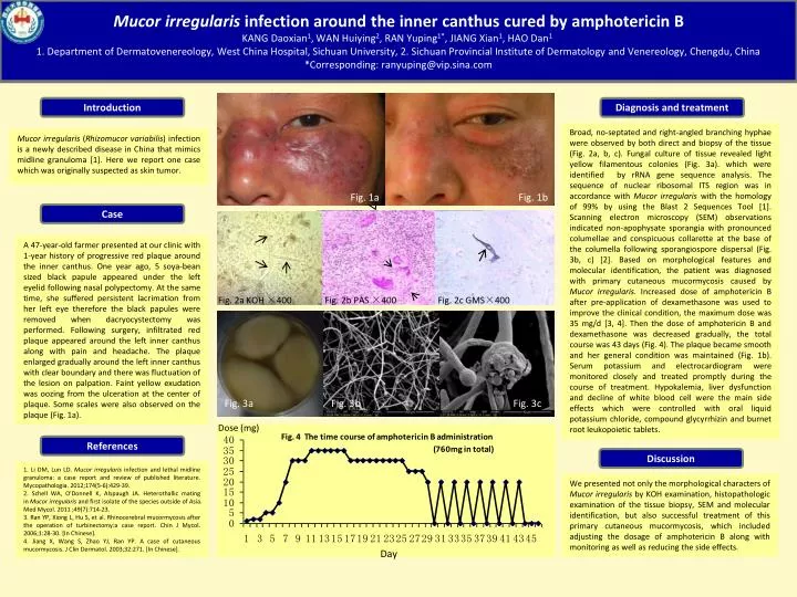
Introduction Diagnosis and treatment Broad, no-septated and right-angled branching hyphae were observed by both direct and biopsy of the tissue (Fig. 2a, b, c). Fungal culture of tissue revealed light yellow filamentous colonies (Fig. 3a). which were identified by rRNA gene sequence analysis. The sequence of nuclear ribosomal ITS region was in accordance with Mucor irregularis with the homology of 99% by using the Blast 2 Sequences Tool [1]. Scanning electron microscopy (SEM) observations indicated non-apophysate sporangia with pronounced columellae and conspicuous collarette at the base of the columella following sporangiospore dispersal (Fig. 3b, c) [2]. Based on morphological features and molecular identification, the patient was diagnosed with primary cutaneous mucormycosis caused by Mucor irregularis. Increased dose of amphotericin B after pre-application of dexamethasone was used to improve the clinical condition, the maximum dose was 35 mg/d [3, 4]. Then the dose of amphotericin B and dexamethasone was decreased gradually, the total course was 43 days (Fig. 4). The plaque became smooth and her general condition was maintained (Fig. 1b). Serum potassium and electrocardiogram were monitored closely and treated promptly during the course of treatment. Hypokalemia, liver dysfunction and decline of white blood cell were the main side effects which were controlled with oral liquid potassium chloride, compound glycyrrhizin and burnet root leukopoietic tablets. Mucor irregularis infection around the inner canthus cured by amphotericin BKANG Daoxian1, WAN Huiying2, RAN Yuping1*, JIANG Xian1, HAO Dan11. Department of Dermatovenereology, West China Hospital, Sichuan University, 2. Sichuan Provincial Institute of Dermatology and Venereology, Chengdu, China *Corresponding: ranyuping@vip.sina.com Fig. 1a Fig. 1b Case A 47-year-old farmer presented at our clinic with 1-year history of progressive red plaque around the inner canthus. One year ago, 5 soya-bean sized black papule appeared under the left eyelid following nasal polypectomy. At the same time, she suffered persistent lacrimation from her left eye therefore the black papules were removed when dacryocystectomy was performed. Following surgery, infiltrated red plaque appeared around the left inner canthus along with pain and headache. The plaque enlarged gradually around the left inner canthus with clear boundary and there was fluctuation of the lesion on palpation. Faint yellow exudation was oozing from the ulceration at the center of plaque. Some scales were also observed on the plaque (Fig. 1a). Fig. 2a KOH ×400 Fig. 2b PAS ×400 Fig. 2c GMS×400 Mucor irregularis (Rhizomucor variabilis) infection is a newly described disease in China that mimics midline granuloma [1]. Here we report one case which was originally suspected as skin tumor. Fig. 3a Fig. 3b Fig. 3c Dose (mg) References Discussion 1. Li DM, Lun LD.Mucor irregularis infection and lethal midline granuloma: a case report and review of published literature. Mycopathologia. 2012;174(5-6):429-39. 2. Schell WA, O'Donnell K, Alspaugh JA. Heterothallic mating in Mucor irregularis and first isolate of the species outside of Asia. Med Mycol. 2011 ;49(7):714-23. 3. Ran YP, Xiong L, Hu S,et al. Rhinocerebral mucormycosis after the operation of turbinectomy:a case report. Chin J Mycol. 2006;1:28-30. [In Chinese]. 4. Jiang X, Wang S, Zhao YJ, Ran YP. A case of cutaneous mucormycosis. J Clin Dermatol. 2003;32:271. [In Chinese]. We presented not only the morphological characters of Mucor irregularis by KOH examination, histopathologic examination of the tissue biopsy, SEM and molecular identification, but also successful treatment of this primary cutaneous mucormycosis, which included adjusting the dosage of amphotericin B along with monitoring as well as reducing the side effects. Day