Download
1 / 32
320 likes | 339 Views
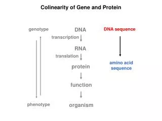
Collinearity of the gene and the protein. http://www.nottingham.ac.uk/bennett-lab/lee.html. As we saw earlier, each gene “specifies” a protein. Therefore, you can’t understand how genes work unless you know some very simple protein biochemistry. Proteins.

E N D
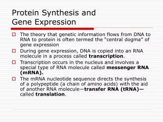
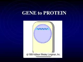
As we saw earlier, each gene “specifies” a protein. Therefore, you can’t understand how genes work unless you know some very simple protein biochemistry. Proteins
Proteins are made by joining together amino acids to form polypeptide chains. Each amino acid in a protein has the same chemical structure except for its “R group”. Proteins
Proteins Amino Acid Contains the following bonded to a central carbon atom: • Amino groups (NH2) • Carboxyl group (COOH) • Hydrogen atom • R group (different in each amino acid) Typical charged in the cell (-NH3+ and COO-)
Proteins 20 different amino acids occur in living cells. 4 chemical groups (composition of the R group): •Acidic (negatively charged), (n = 2) • Basic (positively charged), (n = 3) • Neutral and polar, hydrophilic, (n = 6) • Neutral and non-polar, hydrophobic, (n = 9)
Proteins (Hydrophobic)
Proteins (Hydrophilic)
Proteins Polypeptides N-terminus C-terminus 5’ (DNA) 3’ (DNA) Amino acids are joined to form unbranched polypeptides by a peptide bond
Proteins Proteins show 4 levels of structural organisation: • Primary structure = amino acid sequence • • Determined by the genetic code of the mRNA. • 2. Secondary structure = folding and twisting of a single polypeptide chain. • • Result of weak H-bond and electrostatic interactions. • • e.g., -helix (coiled) and -pleated sheet (zig-zag).
Proteins • 3. Tertiary structure = three dimensional shape (or conformation) of a polypeptide chain. • • Function of R groups contained in the polypeptide. • 4. Quaternary structure = association between polypeptides in multi-subunit proteins (e.g. hemoglobin). • • Occurs only with two or more polypeptides.
When an enzyme carries out a chemical reaction, it is actually the R groups of several of the amino acids that are reacting with the substrate. Polypeptides have to fold up into a particular shape to be functional. It is interactions between the R groups of the amino acids that determine and maintain this shape. Proteins
The first proof of how genes specify proteins came from studies on the oxygen binding protein found in red blood cells: haemoglobin. Haemoglobin is a tetramer. It is made of four polypeptide chains – two -chains and two -chains. Hemoglobin
There are families of people with inherited disorders causing anaemia or thallasaemia. All of the sufferers have altered haemoglobin. These disorders are caused by recessive mutations obeying Mendelian laws. . Hemoglobin Inherited anaemias
One of the best studied is sickle cell anaemia. When the gene defect is in the homozygous form, all of the haemoglobin is altered, the red blood cells become sickle shaped and the sufferers are very ill with severe anaemia. Sickel cell anaemia
Normal blood Sickle cell blood Sickel cell anaemia
In the heterozygous form, only half of the haemoglobin is defective and the anaemia is less severe. Because the blood cells are slightly altered, the heterozygous form confers immunity to malaria. This inherited condition is, therefore, common in parts of West Africa. Sickel cell anaemia
The change in the haemoglobin is very specific. The sixth amino acid is changed from glutamate to valine. This is a change from an acidic, negatively charged, hydrophilic amino acid to a hydrophobic one. Many other inherited anaemias show similarly specific changes of amino acid. Sickel cell anaemia
Why do changes of one amino acid for another destroy the function of a protein? If the protein is an enzyme, the amino acid that carries out the reaction may be changed The altered amino acid may have been involved in pairing with another amino acid to maintain the shape of the protein. Amino acid changes
Sometimes, changing one amino acid for another with very similar properties (e.g. glutamic acid to aspartic acid) might not affect the protein. Mutations in the gene might not change the amino acid – as we will see in the next lecture. Mutations that don’t affect the function of the gene product are called silent mutations. Amino acid changes
The study of haemoglobin has shown that mutations in a gene can cause specific changes in a protein. Different mutations cause different changes. Does the position of the mutation in the gene relate to the position of the changed amino acid in the protein? Collinearity
We can go back to the E. colitrpA cistron to find out the answer. because many mutations in trpA have been mapped and many mutant versions of the TrpA protein have been sequenced to determine the nature and order of the amino acids. Collinearity
Collinearity The tryptophan synthase (trpA) cistron The genetic map and the amino acid sequence are collinear. The mutations in the gene and the changed amino acids in the protein appear in the same relative positions. Positions of mutant loci on genetic recombination map 446 487 223 23 187 58 169 Positions of altered amino acids in protein chain 175 177 183 212 215 234 235
Proteins consist of chains of amino acids and genes consist of chains of nucleotides. - so does each nucleotide specify each amino acid? Collinearity
Collinearity More than one mutation has been found to affect the nature of the amino acid at position 212 in TrpA. Perhaps one nucleotide means glycine, another means arginine and another means glutamate. BUT…
It is possible to get recombination between these two mutants. Therefore, there must be more than one mutable site (presumably more than one nucleotide) specifying each amino acid. Collinearity
Collinearity • In fact, each amino acid is specified by a triplet of nucleotides, known as a codon.
Amino acids and proteins. (2000) In: Instant Notes in Biochemistry. pp 19-42. Hames, B. D. and Hooper, N. M. (Eds). BIOS Scientific Publishers, Oxford Molecular Genetics. (2000) In: An Introduction to Genetic Analysis. pp 271-278. Griffiths, A. J. F,. Miller, J. H., Suzuki, D. T., Lewontin, R. C. and Gelbart, W. M. (Eds). Freeman and Company, New York.