Download
1 / 42
630 likes | 1.21k Views
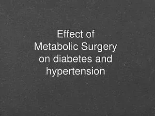
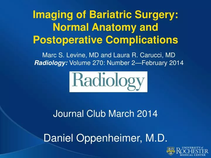
Imaging of Bariatric Surgery: Normal Anatomy and Postoperative Complications. Marc S. Levine, MD and Laura R. Carucci , MD Radiology: Volume 270: Number 2—February 2014 . Journal Club March 2014 Daniel Oppenheimer, M.D. Goals.

E N D
Imaging of Bariatric Surgery:Normal Anatomy and Postoperative Complications Marc S. Levine, MDand Laura R. Carucci, MDRadiology: Volume 270: Number 2—February 2014 Journal Club March 2014 Daniel Oppenheimer, M.D.
Goals • Describe the surgical anatomy and normal imaging findings for three major forms of bariatric surgery • Identify the major complications of these forms of bariatric surgery and their relevant clinical features. • Discuss the imaging findings of bariatric surgery complications on UGI and CT exams
Introduction • BMI (kg/m2) • 25-29 kg/m2 = overweight • 30-35 kg/m2 = obesity • 35+ = morbid obesity • Obesity Epidemic • USA Adults 2004: 66% overweight, 32% Obese • 300,000+ deaths annually • 2nd only to tobacco in preventable deaths • Bariatric Surgery increasing dramatically • 5x # procedures in 2003 vs. 1998
Bariatric Surgery Concepts • Restrictive procedure • Decrease gastric volume early satiety • Laparoscopic adjustable gastric banding and laparascopic sleeve gastrectomy • Bypass procedure • Intentional malabsorption
Laparoscopic Roux-en-Y • Most popular procedure in the USA • Highest long-term success and greater weight loss • Malabsorptive and Restrictive • Stomach divided • Small gastric pouch – restrictive effect • Larger excluded stomach • Jejunum divided • Roux (efferent) limb anastomosed to gastric pouch proximally and jejunojejunostomy distally • Biliopancreatic (afferent) limb anastomosed to jejunojejunostomy http://www.utswmedicine.org
Assessment of Roux-en-Y Bypass • UGI: • Scout image! • Gastric pouch, gastrojejunal anastomosis, Roux limb and jejunojejunal anastomosis • Leak, stricture, obstruction, ulcers • Excluded stomach and biliopancreatic limb not well evaluated • CT: • Oral contrast right before exam • Contrast in gastric pouch, Roux limb to jejunojejunal anastomosis • Excluded stomach collapsed and non-opacified • Leak, stricture, obstruction, ischemia, collections
Roux-en-Y Complications • Leaks • Stricures • Marginal Ulcers • Jejunal ischemia • Small bowel obstruction • Recurrent weight gain
Leak • Up to 5% of patients • ~3/4 at gastrojejunalanastomosis • Usually <10 days post-op • Abscess, peritonitis, sepsis • H20 soluble UGI POD #1-2 • Percutaneous drainage and Abx vs. surgery
Anastomotic Stricture • Transient edema and spasm post op • 4+ weeks post-op • 3-9% of patients • Usually at gastrojejunal anastomosis • Scarring vs. chronic ischemia • Endoscopic dilatation Chandler et. al, AJR 2008;190(1):122–135.
Marginal Ulcers • At gastrojejunal anastomosis • 3-13% of patients • Chronic exposure of gastric acid to Roux limb • Epigastric pain, UGIB • PPIs, surgical revision
Jejunal Ischemia • Acute • Pain, bleeding, N/V early post-op • UGI: Thickened spiculated mucosal folds – submucosal edema and hemorrhage • CT: Bowel wall thickening with mesenteric edema and engorged mesenteric vessecls • Chronic • Intractable N/V secondary to jejunal stricture • UGI: Smooth tubular narrowing, loss of mucosal folds, non-healing giant ulcer(s) > 2.5 cm • CT: Jejunal narrowing with bowel wall thickening
Small Bowel Obstruction • Up to 5% of patients • Adhesions, internal hernias, abd wall hernias, strictures, intussuception • Type A • Dilated Roux limb, decompressed B-P limb and excluded stomach • Type B • Dilated B-P limb and excluded stomach • Closed loop – risk ischemia, perforation • Type C • SBO distal to jejunojejunostomy • Dilated Roux and B-P limbs
Recurrent Weight Gain • Dehiscence of gastric staple line • “Gastrogastric fistula” • Food enters excluded stomach • Restrictive effect gone • Patients no longer have early satiety • Contrast in excluded stomach • Must exclude reflux from B-P limb • Dilation of gastrojejunalanastomasosis another cause recurrent weight gain
Laparoscopic Adjustable Gastric Banding • Silicone band with inflatable balloon sutured around proximal stomach ~2 cm below GE jxn • Creates small gastric pouch • Inflatable inner sleeve – sub q port in abd wall • adjust band intermittently to alter degree of restrictive effect • Early satiety decreased caloric intake • Less invasive vs. Roux-en-Y • Comparable (short-term) weight loss • Fewer complications
Laparoscopic Adjustable Gastric Banding Normal Phi angle 4-58 deg Chandler et. al, AJR 2008;190(1):122–135.
Lap Band Complications • Stomal stenosis • Pouch dilation • Band slippage • Malpositioned Band
Stomal Stenosis • Most common complication, 8-11% • Band overinflation, edema • Excessive luminal narrowing, obstruction • N/V, regurgitation, dysphagia, pain • Findings • Excessive luminal narrowing at band, dilated of proximal stomach/esoph, GE reflux, slow emptying/lack of contrast through stoma • Deflate band +/- repeat fluoro Chandler et. al, AJR 2008;190(1):122–135.
Band Slippage • 4-13% • Band overinflation, recurrent emesis, poor surgical technique • Herniation of fundus above band • Luminal narrowing, obstruction • Increased Phi angle, “O” sign, air-fluid level in gastric pouch • Deflate band +/- surgical correction
Malpositioned Band • Inexperienced surgeon • Band placed in perigastric fat – no restrictive effect • Band placed in lower stomach - gastric outlet obstruction
Perforation • <1% • Traumatic injury to gastric wall at surgery • Pain, fever, leukocytosis • UGI: Contained or free extravasation of contrast • CT: Contrast extravasation, extraluminal gas, fluid collections
Gastric Volvulus • Rare • Band slippage with twisting of stomach around band • Closed loop obstruction • Strangulation, ischemia, infarction • UGI: converging gastric folds, stomach rotated upwards to left above fundus, luminal obstruction • CT: Gastric wall thickening, pneumatosis • Urgent surgical removal
Intraluminal Band Erosion • Late complication, <2% • Pressure necrosis from inflated band • Usually incomplete erosion • Rarely complete erosion • migrate distally in antrum, duodenum, or proximal jejunum or proximally to GE jxn • mechanical obstruction • Contrast surrounding band • Surgical removal
Port and Band-related complications • Port infection and port eversion • Port, tubing or band kink or disruption • Tube erosion into stomach or bowel Chandler et. al, AJR 2008;190(1):122–135.
Port and Band-related complications Chandler et. al, AJR 2008;190(1):122–135.
Laparoscopic Sleeve Gastrectomy • Newer technique - ~5% of bariatric surgeries in 2008 • Stomach divided along long axis • ~75% stomach removed – Banana shaped pouch created – restrictive effect • ~100 cc total volume • No need for periodic adjustments • Irreversible http://www.massgeneral.org Kiriakopolus et. al, Hormones 2009, 8(2):138-143
Laparscopic Sleeve Gastrectomy Complications • Gastric Leak • Gastric Stricture/Gastric outlet obstruction • Gastric Dilation • Gastroesophageal Reflux
Gastric Leak • <1% • Long staple line along greater curvature • Most commonly at proximal end of staple line laterally near GE jxn • Pain, fever, leukocytosis • Extravasation of contrast, extraluminal collections/abscesses
Gastric Stricture/Gastric Outlet Obstruction • Scarring along greater curvature staple line • Narrowing of pouch • Focal strictures or long segment narrowing • Delayed emptying of contrast from residual stomach • Dilated proximal stomach and esophagus • Endoscopic dilatation +/- surgical revision
Gastric Dilation • ~4.5% • Inadequate weight loss, recurrent weight gain • Widening of gastric sleeve, loss of tubular/banana shape
Gastroesophageal Reflux • Increased incidence of GE reflux • up to 20% • Altered gastric anatomy, stasis • Reflux on UGI studies • Esophagitis, Barrett’s esophagus, carcinoma
Question #1 Which of the following anatomic regions usually is NOT opacified with oral contrast material on CT images after Roux-en-Y gastric bypass? a. Gastric pouch b. *Excluded stomach c. Jejunal Roux limb d. Common small bowel channel (distal to jejunojejunostomy)
Question #2 What is the most common site of leak after Roux-en-Y gastric bypass? a. Gastric pouch b. *Gastrojejunalanastomosis c. Blind-ending jejunal stump d. Jejunojejunal anastomosis
Question #3 Which set of findings is most likely to be associated with small bowel obstruction distal to the site of the jejunojejunostomy after Roux-en-Y gastric bypass? a. Collapsed Roux limb and collapsed biliopancreaticlimb b. Dilated Roux limb and collapsed biliopancreaticlimb c. Collapsed Roux limb and dilated biliopancreaticlimb d. *Dilated Roux limb and dilated biliopancreaticlimb
Question #4 7. Which of the following is LEAST likely to be a sign of distal band slippage on abdominal radiographs after laparoscopic adjustable gastric banding? a. Increased Phi angle b. Dilated gastric pouch above band c. *More vertical orientation of band than usual d. O-shaped configuration of band
Question #5 Leaks from the gastric sleeve after sleeve gastrectomymost commonly involve which of the following portions of the gastric staple line? a. *The proximal end of the staple line laterally b. The proximal end of the staple line medially c. The distal end of the staple line laterally d. The distal end of the staple line medially
References • Chandler RC, Srinivas G, Chintapalli KN, Schwesinger WH, Prasad SR. Imaging in bariatric surgery: a guide to postsurgical anatomy and common complications. AJR Am J Roentgenol 2008;190(1):122–135. • Kiriakopoulos A, Varounis C, Tsakayannis D, Linos D. Laparoscopic sleeve gastrectomy in morbidly obese patients. Technique and short term results. Hormones (Athens) 2009; 8: 138-43. • http://www.massgeneral.org • http://www.utswmedicine.org