Download
1 / 62
630 likes | 921 Views
Eyelids & lacrimal apparatus. Eyelid anatomy. The eyelids Protect the eye from injury and excessive light by their closure.

E N D
The eyelids Protect the eye from injury and excessive light by their closure. They also assist in the distribution of tears over the anterior surface of the eye ball. The upper eyelid is larger and more mobile than the lower. The eyelids meet at the medial and lateral angles (or canthi). The palpebral fissure the elliptical opening between the eyelids, is the entrance into the conjunctival sac.
The lateral angle of the eye is directly in contact with the eyeball, whereas themedial rounded angle lies about 6 mm medially from the eyeball. Here the two eyelids are separated by a small triangular space, the locuslacrimalis. in the center of which isa small, pinkish elevation, the caruncula lacrimalis. A semilunar fold, called the plica semilunaris, lies on the lateral side of the caruncle.
Skin: The skin is very thin and easily folds. Microscopic examination of the skin shows many small hairs with sebaceous glands and small sweat glands. The epidermis contains numerous melanocytes. At the margin of the lid the dermis become denser and the papillae are higher.
The eyelashes, are short thick, curved, and more numerouson the upper eyelid (150 in the upper lid and 75 in the lower). They are commonly darker than the scalp hairs, do not becom gray with age, and are replaced every 100 to 150 days. The hair follicles are arranged in two or three rows along the anterior edge of the eyelids and do not possess erector pili muscles, The sebaceous glands of Zeis open into each follicle. Behind and between the follicles modified sweat glands, the ciliary glands of Moll, open into the follicles or onto the eyelid margin. . Subcutaneous tissue: The subcutaneous tissue is very loose and rich in elastic fibers.
Orbicularis OculiThe orbicularis oculi muscle is a flat, elliptical muscle that surrounds the orbital margin extending onto the temporal region and cheek (orbital part); it also (lacrimal portion). It is composed of striated muscle. Beneath the orbicularis oculi muscle lies a thin layer of connective tissue con taining the blood vessels and nerves of the eyelid. Nerve Supply: Temporal and zygomatic branches of the facial nerve enterthe deep surface of the muscle from the lateral side. .
The medial palpebral ligamentattaches the medial ends of the tarsi to the lacrimal crest and the frontal process of the maxilla. The lateral palpebral ligamentattaches the lateral ends of the tarsi to the marginal tubercle on the orbital margin formed by the zygomatic bone. It is a poorly developed ligament. The orbital septum, is perforated by the nerves and blood vessels that exit from the orbital cavityto reach the face and scalp, and by the aponeurotic fibers of the levator palpebrae superioris and the palpebral part of the lacrimal gland. The tarsal gland (meibomian glands) are embedded with in the substance of the tarsal plates. They are arranged in a single row (30 to 40 in the upper lid, and 20_30 in the lower) and the ducts discharge their secretion onto the eyelid margin. When the eyelid is everted, they can be seen as long yellow structures beneath the conjunctiva. The tarsal glands are modified sebaceous glands consisting of a long central canal surrounded by 10 to 15 acini. The mouths of the ducts are lined with stratified squamous epithelium and the cells of the acini are polyhedral cells. The tarsal gland secretion is oily in consistency and prevents the overflow of tears. It also helps to make the closed eyelids airtight. The oily material forms the external layer of the precorneal tear film and hinders rapid evaporation of tears.
Smooth MuscleThe smooth muscle forms the superior and inferior tarsal muscles. The superior tarsal muscle(Muller) is continuous above with the levator palpebrae superioris and below it is attached to the upper edge of the tarsal plate of the upper lid. The function of the superior tarsal muscle is to raise the upper lid and assist the striated muscle of the levator palpebrae superioris. • ConjunctivaThe conjunctiva is a thin mucous membrane that lines the eyelids and is reflected at the superior and inferior fornices onto the anterior surface of the eyeball. It thus covers part of the sclera, and its epithelium is continuous with that of the cornea. At the margin of the eyelid, the conjunctiva continues into the skin along the posterior margin of the openings of the tarsal glands. Here the thinner, nonkeratinized squamous epithelium of the conjunctiva hanges into the keratnized stratified squamous epithelium of the epidermis.
Diseases of the eyelids. • Infections & inflammation of the eyelid glands : e.g. Blepharitis =infection of the eyelid & eyelid margin • Positional abnormalities e.g. ptosis=drooping of the eyelid • Shape abnormalities e.g. entropion=inward deviation of the eyelid, entropion=outward deviation of the eyelid • Miscellaneous e.g. eyelid wart & nevus. • Tumors of the eyelid ,squamous cell & basal cell carcinoma • Trauma & eyelid laceration & primary repair.
Classification of Blepharitis • Anterior Lid Margin • Seborrheic blepharitis • Staphylococcal blepharitis • Mixed seborrheic and staphylococcal blepharitis • Posterior Lid Margin • Meibomian gland dysfunction. • Localized lid margin disease • External hordeolum=Infection of Zeis gland=stye • Internal hordeolum=Infection of meibomian gland • Chalazion=Lipogranulomatous reaction in meibomian gland
Eyelid margin inflammation (blepharitis) is one of the most common problems in ophthalmology. Lid inflammation affects patients of all ages with either an acute or prolonged inflammatory reaction. Despite its frequency,. Treatment is time-consuming and frequently, not completely effective. There are recognized distinctive forms of blepharitis which will be described. Clinically, these diseases have been divided into those which involve mainly the base of the eyelashes (seborrheic blepharitis, staphylococcal blepharitis) and those which involve the meibomian glands (meibomian gland dysfunction). EYELID MARGIN INFLAMMATIONS SEBOHREEIC BLEPHERITIS
SEBORRHEIC BLEPHARITIS • Patients with seborrheic blepharitis complain of continuous burning, itching, light sensitivity, and heaviness of the lids. • Although it can occur at any age, it is frequently found in the elderly. • It is often associated with seborrhea of the scalp, brow, and facial area or of the ears or sternal skin. • Significant findings include eyelid inflammation and dry flakes (dandruff) on the lids (dry seborrheic blepharitis). • A variant of this consists of oily secretions and greasy deposits on the eye lashes (wet seborrheic blepharitis) which may dry to form crusts (scurf). This greasy form may be associated with meibomian gland dysfunction.
With chronicity, some patients develop corneal involvement with a punctate keratopathy in the interpalpebral space. This disease is chronic and incurable. Mild forms can respond to lid hygiene (hot compresses and lid massage with removal of the lid flakes with mild soaps or commercially prepared eye pads). CHRONIC BLEPHERITIS.(EYELID INFECTION)
Patients with Staphylococcal blepharitis often complain of burning, itching, and irritation, especially in the morning They may report difficulty opening their eyes in the morning with their lids matted or stuck together. Treatment ,eyelid hygine, antibiotics drops & ointments as(chloramphenicol,gentamycin,framycetin,tetracycline..) may needed for long time as the condition usually recurrent & chronic. STAPHYLOCOCCAL BLEPHARITIS.
Meibomian gland dysfunction is characterized by bilateral, prolonged, posterior eyelid margin inflammation. Patients complain of redness and burning, presumably from the free fatty acid irritation. Meibomian gland inspissation and occlusion with pouting of the orifices is typical. A thick, yellowish oil can be expressed from individual meibomian glands unless the secretions are inspissated in the orifice MEIBOMIAN GLAND DYSFUNCTION
Painful stye. External hordeolum=Infection of Zeis gland=stye Compared with seborrheic blepharitis patients, patients with S. blepharitis are younger and more frequently female. During acute S. blephararitis, perifolliculitis can lead to ulceration and fibrinous exudates on the lid margin. Typical changes of chronic blepharitis include crusting and hard brittle scales on the base of the lashes. EXTERNAL HORDEOLUM(STYE)
Chalazion • A chalazion is a chronic inflammatory granuloma of a meibomian gland. It appears to be caused by alterations in secretions with retention of secretory material due to obstruction of the ducts. The condition is associated with seborrhea, chronic blepharitis, and acne rosacea. • Chalazia originating in the Zeis sebaceous glands are termed "external Chalazia"; those in the meibomian glands of the tarsus are termed "internal Chalazia." • Clinically, the lesion presents with soft tissue swelling, erythema, and a firm nodule. As the gland fills with oily secretions, it increases in size over weeks.
Patients may develop an acute infection of an occluded meibomian gland (internal hordeolum) or a prolonged obstruction with Lipogranulomatous formation (Chalazia). Chalazia typically erupt under the conjunctival surface, but can also erupt through the tarsus and into the subcutaneous tissue. They may resolve on their own. Occasionally, an incision and drainage or intralesional steroid injections are necessary. With resolution, chalazion frequently result in permanent stellate conjunctival scarring and distortion of the eyelid margin. LONGSTANDING CHALAZION WITH SPONTANEOUS RUPTURE & GRANULOMATOUS CONJUNCTIVAL REACTION
Eyelid hygiene with eyelid massage and expression of meibomian glands should be taught. Ointments should be avoided. Topical corticosteroids are helpful in some of the corneal complications but should generally be avoided. Oral tetracycline or doxycycline are effective in reducing the symptoms associated with meibomian gland dysfunction by an effect on altering the oily products of the meibomian gland. SURGICAL DRAINAGE OF CHALAZION
ECTROPION(OUTWARD) DIRECTED EYELID. • Ectropion is a condition commonly encountered in clinical practice. The pathogenesis of ectropion varies. the evaluation and treatment of the six elements of pathology that may be present in an ectropic eyelid. These factors include: (1) horizontal lid laxity; (2) medial canthal tendon laxity; (3) punctal malposition; (4) vertical tightness of the skin; (5) orbicularis paresis secondary to seventh nerve palsy; and (6) lower eyelid retractor disinsertion.
Entropion is the inurning of the lid margin toward the globe, resulting in corneal irritation from the skin and lashes. Entropion can be classified into three basic groups: congenital, involutional, and cicatricial. the involutional variety remains one of the most common eyelid conditions encountered in practice. The cicatricial variety, although uncommon, is by far the most challenging to treat. ENTROPION(INWARD) DIRECTED EYELID.
Trichiasis & Distichiasis • Trichiasis is defined as normal lashes that have a normal location in the anterior lamella but are misdirected and rub against the cornea and conjunctiva. • Distichiasis also can be an acquired condition, occurring in cases of Stevens-Johnson syndrome, toxic epidermal necrolysis, cicatricial pemphigoid, and chemical and physical injuries of the eyelids. • Patients with distichiasis and trichiasis present with similar symptoms of a watery, red, and irritated eye. • The treatment of distichiasis and trichiasis are similar and based on the extent of lid involvement. If the affected area is limited to a few lashes, simple periodic epilation or electrolysis may suffice. More extensive involvement limited to a localized area of the lid may be treated by focal cryotherapy application.
Blepharoptosis=ptosis= drooping of the upper eyelid • A-Congenital ptosis. • B-Acquired:- • 1-mechanical=heavy eyelid e.g. mass or inflammations • 2-senile=Aponeurogenic Ptosis= disinsertional of the aponeurosis insertion of levator muscle • 3-myogenic=myasthenia gravis • 4-neurogenic=third nerve palsy
Mechanical ptosis may develop secondary to scarring from burn injuries or diseases that can cause severe conjunctival cicatricial changes in the lids such as Stevens-Johnson syndrome or ocular pemphigoid. Large orbital tumors and lid lesions may induce mechanical ptosis. Classic examples include neurofibromas, or hemangiomas of the upper lid MECHANICAL PTOSIS ALLERGY & INFLAMMATION OR TUMORS= “HEAVY EYELIDS” or SECONDARY TO SCARRING
HEREDITARY BLEPHAROPHYMOSIS SYNDROME. • WHAT ARE THE APPARENT ABNORMALITIES???
Blepharoptosis, defined as drooping of the upper eyelid, is a common ophthalmologic condition. The normal upper lid margin rests between 1 to 3 mm below the superior limbus on primary gaze. The approach to a patient with a ptotic eyelid begins with a good history. An important categorical distinction in the evaluation is whether the problem is congenital or acquired. CONGENITAL PTOSIS
Thyroid disease is the Most common cause of unilateral or bilateral lid retraction Surgical treatment of eyelid retraction is usually reserved for patients whose endocrine status and eyelid height have been stable for at least 6 months to 1 year, and in whom retraction causes significant exposure keratopathy, lagophthalmos, chronic conjunctival injection, and cosmetic imperfection. Eyelid retraction
A lid coloboma is a full-thickness developmental defect that may involve the upper eyelid, the lower eyelid, or both. The edges of the defect may be adherent to the bulbar conjunctiva and cornea. CONGENITAL ABNORMALITIES OF THE EYELIDS.COLOBOMA OF THE UPPER EYELID.
Histologically, nevi are classified as junctional, compound, or intradermal depending on the location of the nevus cells in the skin. Most congenital nevi are of compound variety. The "kissing" nevus with mirror-image configuration involving apposed portions of the upper and lower lids is a compound nevus EYELID NAEVUS
Basal cell carcinoma of the eyelid • Basal cell carcinoma is by far the most common malignant tumor involving the ocular adnexa, accounting for 90% of all eyelid malignancies and 20% of all lid tumors. • In the United States, basal cell carcinoma develops in approximately 400,000 people annually!! • The tumor primarily involves the lower lid (50% to 66%). • An incisional biopsy of any suspicious eyelid lesion is required to establish a definitive histologic diagnosis before complete excision and repair.
It constitutes approximately 9% of all periocular cutaneous tumors and is considered the second most common eyelid malignancy. Squamous cell carcinoma of the eyelids is a potentially lethal tumor that can invade the orbit by direct or perineural extension, spread to regional lymph nodes, as well as metastasize to distal sites. Actinic keratoses considered as apremalignant lesions, cutaneous squamous cell carcinoma also may arise from radiation dermatoses, burn scars, and inflammatory lesions Squamous cell carcinoma of the eyelid Is a malignant neoplasm of keratinizing cells of the epidermis.
Malignant melanoma Malignant melanoma represents approximately 5% of all cutaneous cancers. Superficial spreading melanoma is considered the most common variant of melanoma, accounting for 70% of cutaneous melanomas. Clinically, its location on the nonexposed skin surfaces and a more rapid rate of growth are the distinguishing features of this tumor. Malignant melanoma
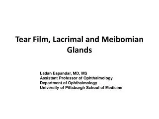
The lacrimal system anatomy & physiology. comprised of three integral components responsible for the production, distribution, and drainage of tears. The main and accessory lacrimal glands secrete a tear film that protects the ocular surface and helps maintain optimal vision. The eyelids and their blinking action help distribute tears across the cornea and transport tears to the puncta. The lacrimal excretory system drains tears from the lacus lacrimalis (Lid tear lake) into the inferior meatus. Conditions altering the complex interplay of anatomy and physiology of these components will result in symptomatic epiphora (tearing). Proper clinical distinction between anatomic and physiologic dysfunction and accurate localization of the anatomic defect are essential for treatment.
The lacrimal system anatomy & physiology. • The main lacrimal gland is located in the superior lateral portion of the anterior orbit. • Directly posterior to the orbital rim is a concavity within the orbital plate of the frontal bone that forms the lacrimal fossa . • The lateral horn of the levator aponeurosis divides the lacrimal gland into an orbital and palpebral lobe, with the palpebral lobe laying beneath the levator aponeurosis. • The orbital lobe contains approximately two thirds of the volume of the lacrimal gland, and the palpebral lobe constitutes the remainder.