Download
1 / 48
480 likes | 638 Views
Protein Secondary Structure. 1958: Kendrew Solves the Structure of Myoglobin.

E N D
1958: Kendrew Solves the Structure of Myoglobin “Perhaps the most remarkable features of the molecule are its complexity and its lack of symmetry. The arrangement seems to be almost totally lacking in the kind of regularities which one instinctively anticipates, and is more complicated than has been predicted by any theory of protein structure”
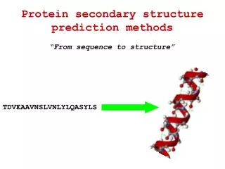
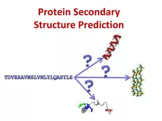
Protein Secondary Structure Protein interior: Hydrophobic core Main chain folds also into interior, but it is highly polar →Problem: Polar atoms must be neutralized through hydrogen bonds →Solution: Regular secondary structure
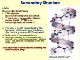
a Helix • Discovered 1951 by Pauling • 5-40 aa long • Average: 10aa • Right handed • Oi-NHi+4 : bb • atoms satisfied • p helix: i - i+5 • 310 helix: i - i+3 1.5Ǻ/res
a Helix is a Dipole … and binds negative charges at N-term
Side Chains project out from the Helix View down one helical turn
CO N C H H2C CH2 CH2 Proline Disrupts Helix No donor!
Frequent Amino Acids at the N-terminus of a helices Ncap, N1, N2, N3 …….Ccap Pro Blocks the continuation of the helix by its side chain Asn, Ser Block the continuation of the helix by hydrogen bonding with the donor (NH) of N3
Helices of Different Character Buried, partially exposed, and exposed
Representation: Helical Wheel Buried, partially exposed, and exposed
Dihedral Angles F and define Backbone Geometry F w The peptide bond w is planar and polar
Ramachandran Plots F All except Glycine Glycine: flexible backbone
Ramachandran Plots F • helix: F, around -60,-50, respectively Other defined regions: b strand and loops
b-Sheet • Involves several regions in sequence • Oi-NHj • Parallel and • anti-parallel • sheets
Antiparallel b-Sheet • Parallel Hbonds • Residue side chains point up/down/up .. • Pleated
Parallel b-Sheet • Less stable than antiparallel sheet • Angled • hbonds
Combined b-Sheet Rare: strains in middle strand
Examples of b-Sheet Topologies Topology diagram Closed barrel
Connecting Elements of Secondary Structure defines Tertiary Structure
Loops • Connect helices and strands • At surface of molecule • More flexible • Contain functional sites
Hairpin Loops (b turns) • Connect strands in antiparallel sheet G,N,D G G S,T
Super Secondary Structures: (1) Greek Key Motif • 24 possible topologies for 2 hairpins • 8 found • Most common: Greek key motif
Super Secondary Structures: (2) b-a-b Motif • Connect strands in parallel sheet
Repeated b-a-b Motif Creates b-meander: TIM Barrel
Protein Classification Alpha contain only a helices Beta contain only b sheets Alpha/Beta contain combination of both Alpha + Beta contain domains of a and b
ALPHA • Occur in • Transmembrane proteins • Structural and motile proteins • Fibrous proteins (Keratin) • Fibrinogen, myosin • Coiled-coils (Leucine Zippers) • 4-helix-bundles • a-helical domains • Globins
ALPHA: Coiled-Coils • Francis Crick, 1953: maximal sc interactions if two helices are wound around each other • Left-handed supercoil: 3.5 residues/turn: • Heptad repeat • “knobs-into-holes” • Leucine zipper motif in Transcription Factors (more about this later..)
ALPHA: 4-Helix Bundle • “ridges-into-grooves” ROP protein
Ridges-into-Grooves • 2 possible arrangements: • i-i+4 ridge: • Globins • i-i+3 ridge: • ROP
ALPHA: a-Helical Domains • >20 a helices form globular domain • Example: muramidase • 27 helices • right-handed • superhelical twist • Hole in center
ALPHA/BETA • Most frequent • 3 classes: • Barrel • Twisted sheet • Horseshoe fold • Functional sites in loop regions
ALPHA/BETA: Barrels • Consecutive a-b-a units • in same orientation • Usually 8; b8-hb- b1 • → closed core of b strands • TIM barrel • Triose Phosphate Isomerase • Usually enzymes
TIM Barrels • aa2,4 point out to helices • branched aasV,I,L • aa1, 3, 5 point into barrel • Bulky hydrophobic aas form tightly packed hydrophobic core • Polar aas (KRE) at tip of barrel: participate in formation of hydrophobic core
TIM Barrels Active site formed by loops at one end of the barrel Distinct from structural region
ALPHA/BETA: Open Sheet • Consecutive a-b-a units • in opposite orientation: • helices on both sides • Rossman Fold • (discovered in 1970 in lactate dehydrogenase) • Many different arrangements
ALPHA/BETA: Horseshoe Fold • Consecutive a-b-a units in same orientation • Not closed: horseshoe • Ribonuclease • Inhibitor • One side points to helix, • The other is exposed
Horseshoe Fold • Leucine-rich repeats • each ~30aa • L responsible for packing
BETA Antiparallel b structures Usually two sheets packed against each other Barrel: composed of anti-parallel strands with hairpin connections Propeller: multi-domain protein
BETA Barrels Retinol-binding protein 8 strands Center: hydrophobic pocket binds lipids
BETA Propellors (I) • Neuraminidase • 6 b-sheets (each 4 strands) organized as propellor blades • Active site formed by loops from each blade • Others: G-proteins, etc
BETA Propellors (II) • Neuraminidase • 6 b-sheets(each 4 strands)organized as propellor blades • Active site formed by loops from each blade
BETA Propellors (III) • Neuraminidase • 6 b-sheets (each 4 strands) organized as propellor blades • Active site formed by loops from each blade
BETA: Jelly-Roll MotifWrapped around a Barrel Composed of repeats of greek keys Concavalin, Hemagglutinin
BETA: b-helix Structures Right-handed coiled structure 18aa: 6 in loop+ 3 in b GGXGXDXUX (U=hydrophobic) Loop stabilized by Ca ion Pectate lyase
Additional Useful Material http://swissmodel.expasy.org/course/text/