Download
1 / 74
1.53k likes | 3.17k Views
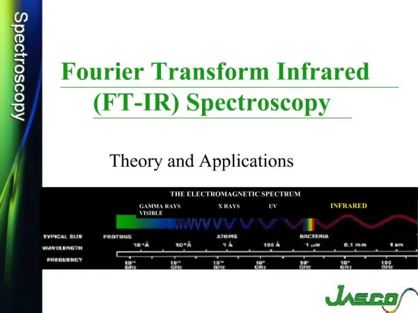
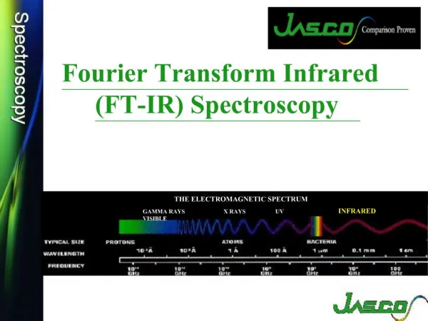
Islamic University in Madinah Department of Chemistry. Infrared Spectroscopy Theory and Interpretation of IR spectra. Prepared By Dr. Khalid Ahmad Shadid. Spectroscopy and the Electromagnetic Spectrum. Basic Theory of IR Absorption.

E N D
Islamic University in Madinah Department of Chemistry Infrared Spectroscopy Theory and Interpretation of IR spectra Prepared By Dr. Khalid Ahmad Shadid
Basic Theory of IR Absorption • Infrared: exciting from one vibrational level to another • UV/Vis: exciting from one electronic level to another • Microwaves: exciting from one rotational level to another
Basic Theory of IR Absorption • Changes in interatomic vibrations of a molecule are brought about through the absorption of IR light
The IR Spectrum • The vibrational spectrum of a molecule is a unique physical property and is characteristic of the molecule • IR spectrum can be used as for identification by the comparison of ‘‘unknown’’ spectrum with reference spectra • IR spectrum can lead to characterization, and possibly even identification of an unknown samples • The IR information can indicate: • Linear or branched backbone • If chains are (un)saturated • Aromatic rings in the structure and substitution • Functional groups
IR Spectroscopy • In IR spectroscopy, there is interaction between molecules and radiations from the IR region of the EMR spectrum (IR region = 4000 - 400 cm-1) • IR radiation causes the excitation of the vibrations of covalent bonds within that molecule. These vibrations include the stretching and bending modes • In practice, it is the polar covalent bonds that are IR "active" and whose excitation can be observed in an IR spectrum • Generally, it is convenient to split an IR spectrum into two approximate regions: • Functional group region: 4000-1000 cm-1 • Fingerprint region:< 1000 cm-1 (more complex and much harder to assign)
Regions of Frequencies Spectral Region Frequency(Hz) Wavenumber(cm-1) Wavelength (,m) After Table 16-1 of Skoog and West, et al. (Chapter 16)
Basic Theory of IR Absorption • We need to talk about • IR energy modes (Types of vibration in molecules) • Band positions • Band Intensity
Infrared Energy Modes • IR energy absorption corresponds to specific modes, corresponding to combinations of atomic movements, such as bending (change in bond angle) and stretching (change in bond length) of bonds between groups of atoms • Energy is characteristic of the atoms in the group and their bonding • Corresponds to vibrations and rotations
Many possible absorptions per molecule exist: stretching, bending,… Vibrational modes leading to IR absorptions:
Bond length changes Symmetrical Stretching Asymmetrical Stretching Bond angle changes Bending: Rocking Scissoring Wagging Twisting تارجحية ذيل الخيل مقصية التوائية او لولبية
Bands Position: Hookes' Law • A vibrating bond In IR can be compared to the physical model of a vibrating spring system that can be described by Hooke's Law of harmonic oscillation • Using the force constant k (which reflects the stiffness of the spring) and the two masses m1 and m2, then the equation indicates how the frequency, u, of the absorption should change as the properties of the system change
Bands Position: Hookes' Law • Hooke’s Law can be used to estimate the wavenumber (n) of light that will be absorbed by chemical bonds • n = 4.12 * (K / m)1/2 K is the force constant (in dynes / cm) and for: single bond: K = 5 x 10⁵ dynes/cm double bond: K = 10 x 105 dynes/cm triple bond: K = 15 x 105 dynes/cm • m = reduced mass • For example in C=C bond: n = 4.12 * (10 x 105 / [12 * 12 / (12 + 12)])1/2 = 1682 cm-1 (calculated) compare to experimental value1650 cm-1
Factors affecting Absorption Frequency • Strength of chemical bond • Masses of attached atoms to the bond • Hydrogen bonding • Resonance • Bond angle or ring strain • Hybridization • Polarity of bonds • External factors: eg. state of measurements, conc., temp., solvent used etc.
Factors Affecting Absorption Frequency: Bond Strength • For a stronger bond (larger k value), u increases Compare (increasing bond strength): • CC bonds: C-C (1000 cm-1), C=C (1600 cm-1) and CºC (2200 cm-1) • CH bonds: C-C-H (2900 cm-1), C=C-H (3100 cm-1) and CºC-H (3300 cm-1) • Strength of the chemical bond: • .
Factors Affecting Absorption Frequency: Masses of Atoms • Masses of the attached atoms to the bond • For heavier atoms (larger m value), u decreases compare (increasing reduced mass): • C-H (3000 cm-1) • C-C (1000 cm-1) • C-Cl (800 cm-1) • C-Br (550 cm-1) • C-I (500 cm-1)
Factors Affecting Absorption Frequency: Hydrogen Bonding • H-bonding • For example: free OH is observed at 3600cm-1 while H-bonded –OH is observed at 3400cm-1
Factors Affecting Absorption Frequency: Resonance • Resonance: electronic factors • Conjugation lowers the energy to vibrate bond • isolated ketones: 1710 cm-1 • a,b-unsaturated ketones: 1690 cm-1 • a,b,g,d-unsaturated ketones: 1675 cm-1
Factors Affecting Absorption Frequency: Bond Strain • Internal factors: Bond angle or ring strain
Factors Affecting Absorption Frequency: Hybridization • Hybridization: • Bonds are stronger in the order sp > sp2 > sp3 • C-H (sp): 3300 cm-1 • C-H (sp2): 3100 cm-1 • C-H (sp3): 2900 cm-1
Factors Affecting Absorption Frequency: Polarity of Bond • The more polar a chemical bond is, the higher the intensity of the band • Low dipole moments results in a weak bands • Low dipole moments results in a weak bands • Band intinsity is qualitatively described as: very strong (vs), strong (s), medium (m), weak (w) and variable (var).
Band intensity • Symmetrical bonds have no dipole moments and thus no IR bands observed in the spectrum (ie. infrared inactive)
Band intensity • Absorption other than fundamental modes of vibration • overtones: exactly 2x or 3x of a fundamental frequency • combination bands where freq. = (1 2). For example: aromatics between 1600 - 2000 cm-1 • coupling: interaction between 2 vibrating groups in close proximity; e.g.:
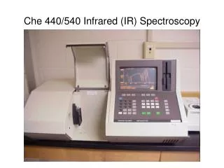
Band intensity • An IR spectrophotometer is made of an IR light source, a sample container, a prism to separate light into different wavelengths, a detector, and a recorder (to produces the infrared spectrum)
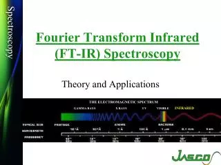
Spectroscopy and the Electromagnetic Spectrum • Radiant energy is proportional to its frequency (cycles/s = Hz) as a wave (Amplitude is its height) • Different types are classified by frequency or wavelength ranges
Infrared Spectroscopy of Organic Molecules • Organic compounds when exposed to electromagnetic radiation, can absorb energy of only certain wavelengths (unit of energy) • Transmits, energy of other wavelengths. • IR region lower energy than visible light (below red – produces heating as with a heat lamp) • 2.5 106 m to 2.5 105 m region used by organic chemists for structural analysis • IR energy in a spectrum is usually measured as wave number (cm-1), the inverse of wavelength and proportional to frequency
4000-2500 cm-1 N-H, C-H, O-H (stretching) 3300-3600 N-H, O-H 3000 C-H 2500-2000 cm-1 CºC and C º N (stretching) 2000-1500 cm-1 double bonds (stretching) C=O 1680-1750 C=C 1640-1680 cm-1 Below 1500 cm-1 “fingerprint” region Regions of the Infrared Spectrum
IR ABSORPTION RANGE The typical IR absorption range for covalent bonds is 600 - 4000 cm-1. The graph shows the regions of the spectrum where the following types of bonds normally absorb. For example a sharp band around 2200-2400 cm-1 would indicate the possible presence of a C-N or a C-C triple bond. Graphics source: Wade, Jr., L.G. Organic Chemistry, 5th ed. Pearson Education Inc., 2003
Differences in Infrared Absorptions • Molecules vibrate and rotate in normal modes, which are combinations of motions (relates to force constants) • Bond stretching dominates higher energy modes • Light objects connected to heavy objects vibrate fastest: C-H, N-H, O-H • For two heavy atoms, stronger bond requires more energy: C º C, C º N > C=C, C=O, C=N > C-C, C-O, C-N, C-halogen
12.8 Infrared Spectra of Hydrocarbons • C-H, C-C, C=C, C º C have characteristic peaks • absence helps rule out C=C or C º C
Infrared Spectra of Some Common Functional Groups • n-Alkanes - look for stretching and bending of C–H and C–C bonds • C–C bends: ca. 500 cm–1 (out of spectral window) • C–C stretches: 1200–800 cm–1, weak bands not of value for interpretation (fingerprint) More characteristic • C–H stretches: occurs from 3000 - 2840 cm–1 CH3: 2962 cm–1, asymmetrical stretch 2872 cm–1, symmetrical stretch CH2: 2926 cm–1, asymmetrical stretch 2853 cm–1, symmetrical stretch • C–H bends: CH3: ca. 1375 cm–1 CH2: ca. 1465 cm–1
n-Alkanes • n-Hexane CH3(CH2)4CH3 CH3 (s) CH3 (as) CH3 (s) C-H Bends CH3 (as) C-H Stretches
Finger printing The IR of C10H22 and C12H26 are Similar but Not Identical C10H22 C12H26
Unconjugated Alkenes • Linear alkenes: • C=C–H stretch: ≥ 3000 cm-1 • C=C–H bending in the range 1000-650 cm-1 • C=C stretch: moderate to weak at 1680-1600 cm-1
Unconjugated Alkenes • Example: 1-Hexene
Cyclic Alkenes • The C=C stretch is sensitive to ring strain (size)
Conjugated alkenes • often conjugation moves C=C stretch to lower frequencies and increases the intensity • The alkene bond stretching vibrations in alkenes without a center of symmetry, e.g. 1-methylbutadiene, gives to two C=C stretches • For symmetrical molecules, e.g. butadiene, only the asymmetric stretch is observed
Conjugated alkenes • 2-methylbutadiene Symmetrical C=C stretch 1640 cm–1 (weak) C-H stretch 3090 cm–1 Out of plane C=C–H bends 990, 892 cm–1 Asymmetrical C=C stretch 1598 cm–1 (strong)
Alkynes • C C–H bend: 700-610 cm-1: broad, strong • C C–H stretch: 3333–3267 cm-1, strong and narrow (as compared to OH or NH) • C Cstretch: weak absorption at 2260-2100 cm-1 • not observed for symmetrical alkynes • terminal alkynes (R-C C-H) absorptions are stronger than internal (R-C C-R) absorptions • Disubstituted or symetrically substituted triple bonds give either no absorption or weak absorption
Terminal Alkynes • Example: 1-octyne Alkyne CC stretch 2119 cm–1 Alkyne C-H bend overtone 1260 cm–1 Alkyne C-H bend 630 cm–1 Alkyne C-H stretch 3310 cm–1
Aromatic Rings • C=C-H stretch occurs at value 3100-3000 cm-1 • C=C-H out of plane bending occurs at 900–690 cm-1 • intense bands, strongly coupled to adjacent hydrogens on the ring • position and number of bands gives information about the ring substitution pattern • C=C stretch occurs in pairs: 1600-1585; 1500-1400 cm-1 • C=C out of plane ring bending: 600-420 cm-1
Aromatic rings • Example: Toluene Overtone bands 2000-1650 cm–1 Aromatic C-H Stretches 3087, 3062, 3026 cm–1 out of plane ring bending 428 cm–1 Aromatic C-H in plane bends 1300-1000 cm–1 Aromatic C-H out of Plane bends 728 cm–1 Aromatic C-C Stretches 1600-1585; 1500-1400 cm–1 694 cm-1