Download
1 / 21
210 likes | 617 Views
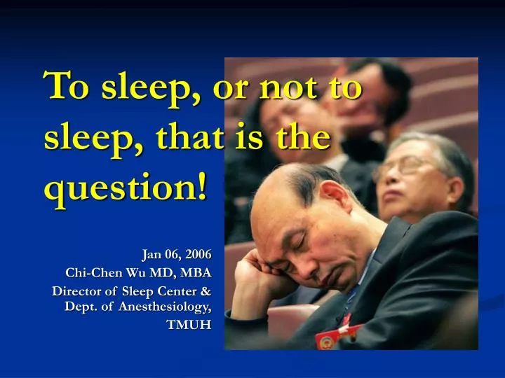
To sleep, or not to sleep, that is the question!. Jan 06, 2006 Chi-Chen Wu MD, MBA Director of Sleep Center & Dept. of Anesthesiology, TMUH. Outlook of sleep centers. Interiors. Hook up, explain, & then sleep!. Test, data collection, scoring. Diagnosis of sleep disorders.

E N D
To sleep, or not to sleep, that is the question! Jan 06, 2006 Chi-Chen Wu MD, MBA Director of Sleep Center & Dept. of Anesthesiology, TMUH
During a sleep study the sleep cycles and stages of sleep are monitored. Electrodes are placed to monitor continuous recordings of brain waves, electrical activity of muscles, eye movement, respiratory rate, blood pressure, blood oxygen saturation, and heart rhythm. Direct observation of the person during sleep may also be used. The test (PSG) is performed for people who suffer from insomnia, excessive daytime sleepiness, obstructive sleep apnea, breathing difficulties during sleep, or behavior disturbances during sleep. 1. Introduction of polysomnography
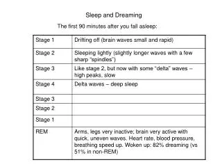
1-1. REM vs. NREM • There are two states of sleep: non-rapid eye movement (NREM) sleep and rapid eye movement (REM) sleep. • REM sleep is associated with dreaming and paralysis of body muscles (except for the eye and diaphragm muscles). • NREM sleep has four stages distinguishable by EEG waves. REM sleep alternates with NREM sleep approximately every 90 minutes. A person with normal sleep usually has four to five cycles of REM and NREM sleep during a night.
1-3. staging of sleep • Sleepstaging was scored according to the criteria of Rechtschaffen and Kales. • Arousals were scored as defined in the American Sleep Disorders Association Atlas Task Force report on EEG arousals. • The arousal index is defined as the number of cortical arousals per hour of sleep (each ≧3 s). • The awakening index is defined as the number of cortical awakenings per hour of sleep (each ≧ 15 s). • The sleep disturbance index is defined as the sum of the arousal index plus the awakening index.
1-4. parameters for staging • Sleep staging depends on : 1. EEG: brain activity 2. EOG: eye movement 3. EMG: muscle tone
1-5-1. Electroencephalogram • Six electrodes (labeled C3, C4, A1, A2 O1, and O2) and one ground electrode are placed around the cranium to record electrical activity across the brain. These leads are used to determine the stage of sleep the patient is in during any given period of the night.
1-5-2. Electroocculogram • One electrode is placed above and to the outside of the right eye, and another electrode is placed below and to the outside of the left eye. • These leads record the movements of the eyes during sleep and serve to help determine sleep stages.
1-5-3. Electromyogram • Three leads are placed on the chin (one in the front and center and the other two underneath and on the jawbone) and two are placed on the inside of each calf muscle 2-4cm apart. • These leads serve to demonstrate muscle movement during sleep. This is helpful in documenting a wake period, an arousal, or just a spastic movement.
1-5-4. ECG and flow detector • Two electrodes are placed on the upper chest near the right and left arms. These record the heart rate and rhythm and serve to alert the technician to a possible emergency situation. They also demonstrate whether apneic desaturation leads to arrhythmias or not. • It senses the amount of air moving into and out of the airways and sends a signal to a physiological recorder. This tracing is used to determine the presence and extent of apneic episodes.
1-5-5. Pulse oximetry • The O2 saturation is measured by a pulse oximeter probe placed on the patient i.e. finger, earlobe, etc.
1-5-6. Respiratory Effort • Two Velcro bands, one placed around the chest under the breasts and one around the abdomen, serve to determine chest wall and abdominal movements during breathing. Each band is joined together by a piezo crystal transducer. • The force of chest/abdominal expansion on the bands stretches the transducer and alters the signal to a physiological recorder. These leads, combined with the airflow sensor, are how apnea is demonstrated and categorized during the test.
1-5-7. Video recording • If the sleep disorders center is equipped with video cameras in the patient rooms, the patient can be taped while sleeping. This allows the technician to review the tape at any time during the test and verify whether strange looking waveforms were caused by an actual arousal, a period of wake, or normal patient movement in bed.
1-5-9. Instruments for sleep study in 1982 Dr. William C. Dement, a leading authority on sleep disorders, posed in 1982 next to an EEG machine, a sleep research tool for studying eye movement, muscle tension and brain waves. Photo: Stanford News Service archives