Download
1 / 24
250 likes | 422 Views
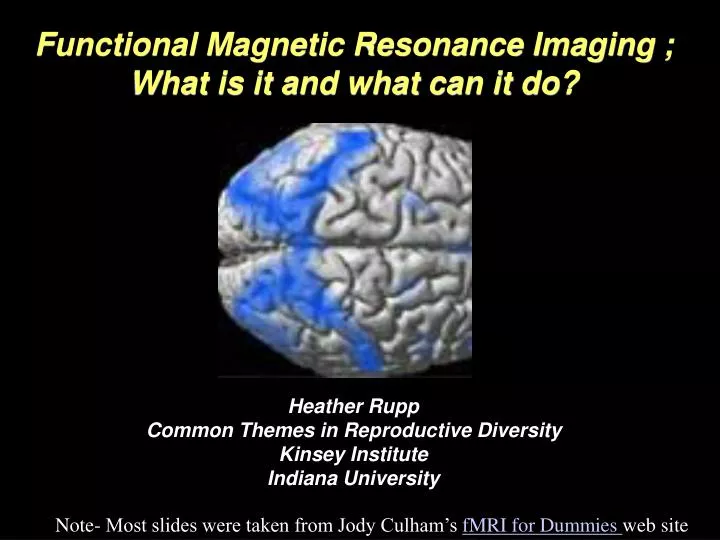
Functional Magnetic Resonance Imaging ; What is it and what can it do?. Heather Rupp Common Themes in Reproductive Diversity Kinsey Institute Indiana University. Note- Most slides were taken from Jody Culham’s fMRI for Dummies web site. Today. I. Structural Scans II. Functional Scan

E N D
Functional Magnetic Resonance Imaging ; What is it and what can it do? Heather Rupp Common Themes in Reproductive Diversity Kinsey Institute Indiana University Note- Most slides were taken from Jody Culham’s fMRI for Dummies web site
Today I. Structural Scans II. Functional Scan III. Simulator IV. Study Design
Today I. Structural Scans II. Functional Scan III. Simulator IV. Study Design
Structural • Different Pulse Sequences (T1, T2, EPI) • Different Views • Think about how to optimize scans for certain brain regions that may interest you
T1 and TR • T1 = recovery of longitudinal (B0) magnetization • used in anatomical images • ~500-1000 msec (longer with bigger B0) • TR (repetition time) = time to wait after excitation before sampling T1 Source: Mark Cohen’s web slides
T2 and TE T2 = decay of transverse magnetization TE (time to echo) = time to wait to measure T2 or T2* (after refocusing with spin echo or gradient echo) Source: Mark Cohen’s web slides
Today I. Structural Scans II. Functional Scan III. Simulator IV. Study Design
Today I. Structural Scans II. Functional Scan III. Simulator IV. Study Design
How Attractive? 1 = Very Unattractive 2 = Unattractive 3 = Attractive 4 = Very Attractive
Today I. Structural Scans II. Functional Scan III. Simulator IV. Study Design
Cognitive subtraction originated with reaction time experiments (F. C. Donders, a Dutch physiologist). Measure the time for a process to occur by comparing two reaction times, one which has the same components as the other + the process of interest. Subtraction Logic Example: T1: Hit a button when you see a light T2: Hit a button when the light is green but not red T3: Hit the left button when the light is green and the right button when the light is red T2 – T1 = time to make discrimination between light color T3 – T2 = time to make a decision Assumption of pure insertion: You can insert a component process into a task without disrupting the other components. Widely criticized
Change only one thing between conditions! • Two paired conditions should differ by the inclusion/exclusion of a single mental process • How do we control the mental operations that subjects carry out in the scanner? • Manipulate the stimulus • works best for automatic mental processes • Manipulate the task • works best for controlled mental processes • DON’T DO BOTH AT ONCE!!! Source: Nancy Kanwisher
Add a “one back” task • subject must hit a button whenever a stimulus repeats • the repetition detection is much harder for the scrambled shapes • any activation for the intact shapes cannot be due only to attention Time Dealing with Attentional Confounds fMRI data seem highly susceptible to the amount of attention drawn to the stimulus or devoted to the task. How can you ensure that activation is not simply due to an attentional confound? Add an attentional requirement to all stimuli or tasks. • Other common confounds that reviewers love to hate: • eye movements • motor movements
Blocked Design Block Designs = trial of one type (e.g., face image) = trial of another type (e.g., place image) Assumption: Because the hemodynamic response delays and blurs the response to activation, the temporal resolution of fMRI is limited. WRONG!!!!!
Blocked vs. Event-related Source: Buckner 1998
Thought Experiments • What do you hope to find? • What would that tell you about the cognitive process involved? • Would it add anything to what is already known from other techniques? • Could the same question be asked more easily & cheaply with other techniques? • Would fMRI add enough to justify the immense expense and effort? • What would be the alternative outcomes (and/or null hypothesis)? • Or is there not really any plausible alternative (in which case the experiment may not be worth doing)? • If the alternative outcome occurred, would the study still be interesting? • If the alternative outcome is not interesting, is the hoped-for outcome likely enough to justify the attempt? • What would the headline be if it worked? • What are the possible confounds? • Can you control for those confounds? • Has the experiment already been done?
Top Ten Things Sex and Brain Imaging Have in Common 10. It's not how big the region is, it's what you do with it. 9. Both involve heavy PETting. 8. It's important to select regions of interest. 7. Experts agree that timing is critical. 6. Both require correction for motion. 5. Experimentation is everything. 4. You often can't get access when you need it. 3. You always hope for multiple activations. 2. Both make a lot of noise. 1. Both are better when the assumption of pure insertion is met. Source: students in the Dartmouth McPew Summer Institute