Download
1 / 1
10 likes | 186 Views
OH. NH 2. NH 2. NH 2. NH 2. OH. OH. OH. OH. NH 2. OH. NH 2. OH. NH 2. OH. NH 2. NH 2. OH. PDMS wells on substrate. AFM Image of the mini-well edge after Neutravidin deposition. Mini-Well Array Assembly. PDMS block. Silicon chip. array chip. UV Exposure. 1. Biotin-Si.

E N D
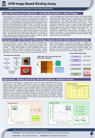
OH NH2 NH2 NH2 NH2 OH OH OH OH NH2 OH NH2 OH NH2 OH NH2 NH2 OH PDMS wells on substrate AFM Image of the mini-well edge after Neutravidin deposition. Mini-Well Array Assembly PDMS block Silicon chip array chip UV Exposure 1 Biotin-Si Neutravidin Fiducial marks Punch wells APTES vapor deposition Array Chip 2 Plasma treat Biotinylate Si step height = 3.1 nm NHS-LC-LC-biotin 3 Place PDMS on silicon chip Neutravidin deposition 4 Biotinylated-HS deposition 5 Binding Specificity Serotype Density of AAV on Substrate Surface (# of particles/mm) Neutravidin bHS AAV1 0.18 ± 0.05 0.06 ± 0.03 AAV2 0.42 ± 0.06 15.98 ± 1.01 AAV3 0.64 ± 0.06 31.48 ± 1.91 AAV5 <0.00 <0.00 = de-OS heparin = de-6-OS heparin = de-NS heparin = de-2-OS heparin virus density (particles/25 mm2) Log (inhibitor concentration) (mM) AFM Image-Based Binding Assay CISMM: Computer Integrated Systems for Microscopy and Manipulation Project Motivations and Objectives: Viral Binding Studies for Gene Therapy Viral infections rely on the virion particle binding to cell surface receptors. Understanding the mechanisms involved in this interaction is relevant in research fields such as gene therapy, drug development and delivery, and biosensors. In collaboration with UNC Gene Therapy Center and School of Pharmacy, we are specifically investigating the interaction between human parvovirus adeno-associated virus type 2 (AAV2), a potential viral vector, and heparan sulfate proteoglycan (HSPG), a cell surface receptor for AAV2. AAV is a highly attractive gene therapy vector due to its stability, low toxicity, low immunogenecity and ease in large scale production. AAV has been identified as a potential vector to treat numerous diseases such as brain tumors, muscular dystrophy, hemophilia and cystic fibrosis. A limitation of AAV is its broad host range which makes treatment of specific tissues difficult. Research efforts have been directed to target the vectors to specific cell types by modifiying the binding sites on the capsid. Further understanding of the interactions between AAV capsid proteins and heparan sulfate (HS) will aid in the modification of the AAV for tissue specific infectivity. Heparan sulfate is a polysaccharide chain of sulfated iduronic/glucuronic and sulfate glucosamine residues and can be differentiated into sequence specific structures by enzymes. These enzymatic modifications, known as heparan sulfate sulfotransferase modifications, have been shown to change the biological nature of heparan sulfate such as specific binding to proteins and viruses. For understanding HS-assisted viral infection mechanisms, we are interested in investigating the binding affinity and stability of AAV to different HS structures. We have developed a model heparan sulfate surface in which AAV adsorption studies are done and analyzed using the atomic force microscope (AFM). Development: Mini-Well Array Methodology, Heparan Sulfate Surface Functionalization We have developed an assay system which utilizes photolithography, functionalized silicon surfaces, PDMS mini-wells and atomic force microscope (AFM) imaging to study the specific interactions between HS and AAV. Viruses were adsorbed on to HS functionalized surface and virus receptor binding is directly measured using the AFM. Another advantage of using the AFM is its ability to simultaneously provide structural and quantitative information about the binding interaction at the single molecule level in a controlled environment. In addition, the system has the flexibility to independently study different viruses binding to the same receptor, the same virus binding to different receptors, or use soluble molecules to compete with the virus-receptor binding. This technique has been optimized to minimize reagent use (reaction volumes can be as small as 1 ml) and to facilitate analysis. Surface Modification Steps Experiments: Binding Specificity, Binding Isotherms, Inhibition Studies The AFM-based methodology is currently used to study the binding interaction between adeno-associated virus type 2 (AAV2), a potential viral vector, and heparan sulfate (HS), an AAV2 cell surface receptor. Adsorption of AAV2 on to the functionalized surfaces are analyzed using the AFM. Binding of different AAV serotypes on the HS-modified surface confirmed that AAV2 and AAV3 have significant HS binding compared to AAV1 and AAV5. Isotherms are performed to determine binding constants of proteins. A binding constant of 340pM was determined for AAV2 to HS. IC50s of relevant molecules (such as chemically de-sulfated heparin) can be determined by performing inhibition studies in the reaction wells and using AFM to analyze the variance in the virus adsorption density. Preliminary results indicate a strong interaction between de-2-OS heparin and AAV2. Inhibition studies can lead to discovering potential anti-viral drugs as well provide structural information about the number of binding sites on the capsid. Inhibition Studies Binding Isotherm virus density (particle/mm2) Note: lines are only guides for the eye virus concentration (nM) http://cs.unc.edu/Research/nano/cismm/biophysics Collaborators: Jude Samulski (UNC Gene Therapy Center), Doug McCarty (UNC Gene Therapy Center) Project Lead: Jian Liu (Schooll of Pharmacy) Investigators: Atsuko Negishi, Richard Superfine 11.21.03