Download
1 / 75
750 likes | 752 Views
Learn about the different cell types of neural tissue, including neurons and neuroglial cells. Explore the structure and function of neurons, and understand how neuroglia support and nourish neurons. Discover the divisions of the nervous system and their functions.

E N D
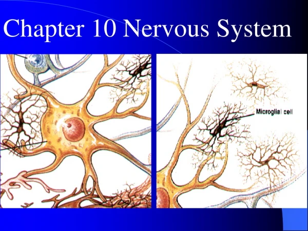
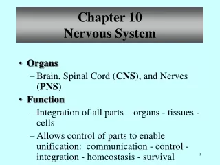
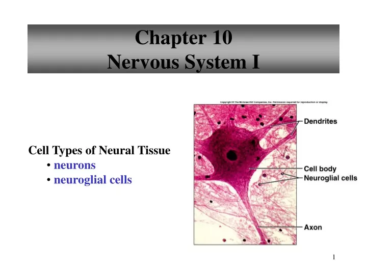
Chapter 10Nervous System I Cell Types of Neural Tissue • neurons • neuroglial cells
Composed of some blood vessels and connective tissue but mostly neural tissue (2 cell types) neurons neuroglia • Neurons transmit information as nerve impulses along nerve fibers • Nerves are bundles of nerve fibers
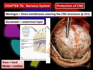
Neuroglia surround, support, and nourish neurons and might even send and receive messages • Synapses are spaces between neurons • Neurotransmitters are biological messengers • Central nervous system (CNS) consists of the brain and spinal cord
Peripheral nervous system (PNS) consists of the peripheral nerves that connect the CNS to other body parts • CNS and PNS provide 3 general functions: sensory, integrative, and motor
Sensory receptors at the ends of peripheral neurons gather information: Info nerve impulse CNS integration decision made motor neurons muscles or glands (effectors)
Divisions of the Nervous System • Central Nervous System • brain • spinal cord • Peripheral Nervous System • nerves • cranial nerves • spinal nerves
Divisions of Peripheral Nervous System Sensory Division • picks up sensory information and delivers it to the CNS Motor Division • carries information to muscles and glands Divisions of the Motor Division • Somatic – carries information to skeletal muscle • Autonomic – carries information to smooth muscle, cardiac muscle, and glands
Functions of Nervous System Sensory Function • sensory receptors gather information • information is carried to the CNS Motor Function • decisions are acted upon • impulses are carried to effectors Integrative Function • sensory information used to create • sensations • memory • thoughts • decisions
Mature neurons generally do not reproduce • All neurons have a cell body and nerve fibers • Cell body contains: granular cytoplasm, mitochondria, lysosomes, a Golgi apparatus, microtubules • Neurofibrils (fine threads) extends into and supports fibers
Chromatophilic substance (nissl bodies) made of rough endoplasmic reticulum is scattered in cytoplasm • Cytoplasmic inclusions include glycogen, lipids, and pigments • Large nucleus near the center with a nucleolus • Nerve fibers, dendrites and axons extend from the cell body
Dendrites are usually branched and communicate with other neurons • Axons carry nerve impulses away from the cell body • Axons also convey biochemicals produced in the cell body (axonal transport) • Schwann cells wind around axons in layers called myelin
Myelin has higher lipids than other surface membranes and forms a myelin sheath on the outside of axons • A neurilemmal sheath made of Schwann cells that have most of the cytoplasm and nuclei forms outside the myelin sheath • Nodes of Ranvier are gaps in myelin sheath between Schwann cells
Myelinated fibers appear white (white matter in brain and spinal cord) • Small axons don’t have myelin (unmyelinated) and appears gray (gray matter in brain and spinal cord)
Myelination of Axons White Matter • contains myelinated axons Gray Matter • contains unmyelinated structures • cell bodies, dendrites
Classification of Neurons – Structural Differences Bipolar • two processes • eyes, ears, nose Unipolar • one process • ganglia Multipolar • many processes • most neurons of CNS
Neurons • Vary in size and shape • May differ in length and size of axons and dendrites • Vary in the number of processes by which they communicate with other neurons • Vary in function
3 types classified by structure: bipolar, unipolar, multipolar (Table 10.1) • 3 types classified by function: sensory, interneuron, motor (Table 10.1)
Classification of Neurons – Functional Differences Sensory Neurons • afferent • carry impulse to CNS • most are unipolar • some are bipolar Interneurons • link neurons • multipolar • in CNS Motor Neurons • multipolar • carry impulses away from CNS • carry impulses to effectors
Types of Neuroglial Cellsin the PNS Schwann Cells • produce myelin found on peripheral myelinated neurons • speed neurotransmission Satellite Cells • support clusters of neuron cell bodies (ganglia)
Neuroglia • Guide neurons in the embryo to their positions and may stimulate them to specialize • Produce growth factors that nourish neurons • Remove ions and neurotransmitters that build up in between neurons, allowing continued information transmission
Some may communicate with neurons • Schwann cells are the neuroglia of the PNS • CNS neuroglia: astrocytes, oligodendrocytes, microglia, and ependyma
Neuroglia form more than half the volume of the brain • Most brain tumors are neuroglia that multiply too often
Types of Neuroglial Cellsin the CNS Astrocytes • CNS • scar tissue • mop up excess ions, etc • induce synapse formation • connect neurons to blood vessels Microglia • CNS • phagocytic cell Ependyma • CNS • ciliated • line central canal of spinal cord • line ventricles of brain Oligodendrocytes • CNS • myelinating cell
Mature neurons do not reproduce • Injury to a neuron cell body usually kills it • Damaged axons in a peripheral nerve may regenerate (3-4mm /day), but may end up in the wrong place so full function often does not return
Neuroglial cells assist in regeneration (nerve growth factors) • Damaged axons in the CNS are unable to produce myelin and regeneration is unlikely
The Synapse Nerve impulses pass from neuron to neuron at synapses
Synaptic Transmission Neurotransmitters are released when impulse reaches synaptic knob
Cell Membrane Potential • A cell membrane is polarized (electrically charged) due to unequal distribution of ions. The inside is negative with respect to the outside.
Distribution of Ions • K+ are the major ions inside the cell (intracellular) • Na+ are the major extracellular ions • Channels in the membrane allow movement in and out • Chemical and electrical factors affect the opening and closing of these gatelike channels
Resting nerve cell is not being stimulated to send an impulse • K+ pass through resting cell membranes much easier than Na+ • Ca2+ less able to cross a resting cell membrane than K+ or Na+
Resting Membrane Potential • inside is negative relative to the outside • polarized membrane • due to distribution of ions • Na+/K+ pump
Active transport keeps a greater concentration of K+ inside the cell and a greater concentration of Na+ outside the cell • Cytoplasm contains anions: (PO4-2), (SO4-2), and proteins that can’t diffuse through the cell membrane
Na+ and K+ follow the laws of diffusion (high to low) • more K+ diffuses out than Na+ can move in • more + charges leave the cell than enter • outside of the cell has a positive charge and the inside has a negative charge
Difference in electrical charge = potential difference (measured in volts) • Difference between inside and outside = -70 millivolts • Resting potential = separation of charge • Action potential = work it may do to send a nerve impulse
Local Potential Changes • caused by various stimuli • temperature changes • light • pressure • environmental changes affect the membrane potential by opening a gated ion channel
Local Potential Changes • if membrane potential becomes more negative, it has hyperpolarized • if membrane potential becomes less negative, it has depolarized • graded • summation can lead to threshold stimulus that starts an action potential
Nerve cells can respond to changes in their surroundings • Changes affect the resting potential of the membrane • Hyperpolarized = membrane potential becomes more negative than resting potential
Depolarized = membrane potential becomes more positive than resting potential (Na+ move in) • Threshold potential = strong enough depolarization to conduct a nerve impulse (action potential)
Action Potentials • at rest membrane is polarized • threshold stimulus reached • sodium channels open and membrane depolarizes • potassium leaves cytoplasm and membrane repolarizes
Action Potentials • Reached when the membrane potential becomes positive • When the threshold potential is reached Na+ move into the cell causing the membrane potential to become more positive (as high as +30 mV) depolarization
At the same time K channels open and allow K+ to move out • causing the inside to become negative again repolarization • Rapid sequence of depolarization and repolarization causes an electric current to move down the nerve fiber nerve impulse