Download
1 / 29
300 likes | 491 Views
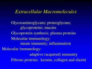
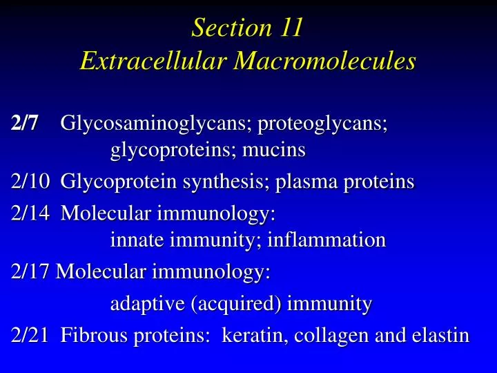
Section 11 Extracellular Macromolecules. 2/7 Glycosaminoglycans; proteoglycans; glycoproteins; mucins 2/10 Glycoprotein synthesis; plasma proteins 2/14 Molecular immunology: innate immunity; inflammation 2/17 Molecular immunology: adaptive (acquired) immunity

E N D
Section 11 Extracellular Macromolecules 2/7 Glycosaminoglycans; proteoglycans; glycoproteins; mucins 2/10 Glycoprotein synthesis; plasma proteins 2/14 Molecular immunology: innate immunity; inflammation 2/17 Molecular immunology: adaptive (acquired) immunity 2/21 Fibrous proteins: keratin, collagen and elastin
Section 11 Extracellular Macromolecules 1. Glycosaminoglycans Proteoglycans GlycoproteinsMucins 2/7/06
Extracellular Macromolecules macromolecule% carb. glycosaminoglycans* (GAGs) 100 proteoglycans* 90-95 glycoproteins 2-30 fibrous proteins 1-2 Examples of functions: mechanical support lubrication cushioning adhesives cell spacers selective filters * aka mucopolysaccharides, mucoproteins, respectively 1
Extracellular matrix in tissues • ground substance + fibers • macromolecules between cells • ground substance molecules GAGs/proteoglycans (mostly carbohydrate) • fibers fibrous proteins: structural adhesive • especially abundantin connective tissue epithelial cells adhesionmolecules extra-cellularmatrix basallamina underlying cells 2 Adapted from Hypercell
GAG structure A sugar • exist as: • independent moleculese.g., hyaluronate & heparin • parts of larger structurese.g., in proteoglycans • heteropolysaccharides repeating structure: disaccharide (AB)nABABAB… • where A is usually 1 uronic acid (hexose with C6 as COO– ) • & B is 1 glycosamine (amino sugar) derivative • unbranched • glycosidic linkage • anomeric C of 1 unit linked to hydroxyl of adjacent unit B sugar 3
GAG structure: repeating units 4 GAG A sugar B sugarhyaluronate glucuronate N-acetyl glucosamine * 2 5
GAG structure: repeating units 4 GAG A sugar B sugarhyaluronate glucuronate N-acetyl glucosamine chondroitin sulfate glucuronate N-Ac galactosamine 4-SO4 dermatan sulfate iduronate " heparan sulfate glucuronate glucosamine N-SO3, 6-SO4 heparin iduronate 2-SO4 " keratan sulfate galactose N-Ac glucosamine 6-SO4 *opposite configuration in iduronate glucuronate/iduronate: epimers at C5 glucose/galactose: epimers at C4 * 2 5
Hyaluronate (aka hyaluronan) 5 • mol wt: 106 – 107 (5000 – 50,000 monosaccharide units) • very polar: 2 hydroxyls/unit 6 heteroatoms/unit COO– every other unit binds cations: Na+, Ca++ Display of HA in motion A B A B A B hyastk2.gif – – – 1 2 3 4 5 6 (glucuronate–N-acetyl glucosamine)3 (glcUA–glcNAc)3
Hyaluronate: structure & properties 6 • extended structure (charge repulsion) • hydrophilic: binds 10–100 × wt in H2O • additional, loosely associated H2O, so that volume occupied ~1000 × weight Display of HA with glcUAs in CPK hyacpk2.gif – – – 2 3 1 4 6 5 (glcUA–glcNAc)3 glcUAs in space-filling form (CPK)
Hyaluronate Alberts et al. Fig. 19-37 • solutions viscous, gel–like, compression-resistant • occurrence: EC matrix,esp. in developing tissue healing wounds synovial fluid • functions: lubricant shock absorber flexible cement attachment site path for cell migration • made by fibroblasts • degraded by hyaluronidase hyaluronidase • bacterial hyaluronidase facilitates spread of infection 7
Heparin • mol wt ~ 104 • ~ 40 monosaccharide units • made & released from mast cells in lungs & liver heparin cell 8
Heparin • mol wt ~ 104 • ~ 40 monosaccharide units • made & released from mast cells in lungs & liver • also associated with luminal surface of endothelium • anticoagulant • forms complex with antithrombin III • this complex binds to thrombin, inactivating it • as a result, clot growth is limited • fast-acting, making it therapeutically useful heparin cell 8
Extracellular Macromolecules macromolecule% carb. glycosaminoglycans* (GAGs) 100 proteoglycans* 90-95 glycoproteins 2-30 fibrous proteins 1-2 Examples of functions: mechanical support lubrication cushioning adhesives cell spacers selective filters * aka mucopolysaccharides, mucoproteins, respectively
Proteoglycans (PGs) • composed of as many as 200 GAG chains covalently bonded to a core protein via serine side chains • molecular weight range: 105 – 107 • GAG chains: chondroitin sulfate, heparan sulfate, dermatan sulfate, keratan sulfate Examples • decorin • many connective tissues • binds type I collagen, TGF-b • perlecan • basal laminae • structural & filtering function • aggrecan • syndecan (slide 13) from Alberts et al. Fig. 19-57 AlbertsT 19-3:Dcrn GAGchndSO4/drmSO4 GAG chains core protein 9
PG in basal lamina of renal glomerulus adapted from Alberts et al., 3 ed., Fig. 19-56 • network offibrousproteins &perlecanPG forms filter entactin perlecan GAG:heparan SO4 laminin type IV collagen 10
Proteoglycans: aggrecan • ~100 GAG chains/molecule • ~100 monosaccharides/GAG chain • each "bristle" = 1 GAG chain • each GAG chain is either chondroitin sulfate or keratan sulfate • GAG chains linked to ser side chains of core protein based onAlberts et al. Fig. 19-37 4ed. 19-40 core protein GAG chains 11
An aggregate of aggrecans & hyaluronan 1m • major GAG–PGin cartilage • link proteins bind noncovalently • with bound H2O,disperses shocks,compressive force • ~ cell size • adhesion proteins link to collagen & cells • degraded by chondroitin sulfatase, etc core protein link proteins hyalur-onan keratansulfate chondroitinsulfate 12 Alberts et al. Fig. 19-41
Proteoglycans: syndecan • cell-surface PG • core protein domains • intracellular • transmembrane • extracellular 5 GAGs attached • functions • interactions • cell-cell • cell-matrix • growth factor receptor GAG chains outside inside core protein Lehninger et al.Fig. 9-22 13
GAG synthesis & breakdown –UDP • synthesis • activated precursors: UDP–monosaccharide derivativese.g., UDP–glucuronate • residues added one at a time in Golgi complex • sulfate moieties • donor: PAPS (active sulfate) • degradation • lysosomes • specific glycosidases & sulfatases • mucopolysaccharidoses • genetic disorders • accumulation of GAG due to absence of a specific glycosidase or sulfatase – adenine – – – 14
Extracellular Macromolecules macromolecule% carb. glycosaminoglycans (GAGs) 100 proteoglycans 90-95 glycoproteins* 2-30 fibrous proteins 1-2 * polypeptide with 1 or more oligosaccharide side chains 15
Glycoproteins: functions of glyco moieties • increase protein’s solubility & hydrophilicity (sl 19) • stabilize protein against • denaturation • proteolysis • markers • direct protein's destination • organelle • plasma membrane • export (secretion) • indicate protein's lifetime (sl 21) • part of the protein's receptor recognition site (sl 23) • signal factors such as hormones, cytokines • cell-cell adhesion proteins Glycosylation:one kind of post-translational modificationothers: phosphorylation carboxylation 16
Glycoprotein structure 17 • polypeptide with 1 or more oligosaccharide side chains • oligosaccharide linked to polypeptide in two ways: type linked to side chain of organelle where sugars are added to protein O-linked serine (ser), threonine (thr), Golgi complex lumen (O-glycoside) hydroxylysine (in collagen) N-linked asparagine (asn) rough ER lumen (N-glycoside)
Glyco moiety structure • oligosaccharide chain extends away from protein surface • units mostly hexoses in pyranose (6-atom ring) form • branched • glycosidic links varied: or 1,2; 1,3; 1,4 • terminal sugaroften sialate 2 asn 7 2 7 asn 18 Stryer 4ed., p. 463
Mucins: salivary glycoproteins • mol wt ~ 106 • ~800 short (disaccharide) side chains • terminal sugar is sialate • anionic sugar • at end of glyco chains of many glycoproteins • very hydrophilic, extended structure ~ ~ galNAc sialate – 2 19
Mucins: modification & aggregation ~ ~ ~ ~ • sialidase (neuraminidase) • catalyzes hydrolysis of sialates from mucins • secreted by oral bacteria • products: • less hydrophilic, less H2O-soluble, more folded, more aggregated • part of the enamel pellicle & dental plaque matrix ~ ~ galNAc sialate x H2O sialidase x 20
Role of glyco moiety in controlling protein lifetime • many blood proteins have glyco chains with terminal sialate • endothelial surface sialidases slowly remove sialates from these circulating proteins • rate of sialate removal depends on protein's structure • now-exposed gal–glcNAc… residues bind to asialoglycoprotein receptor on liver cell surface • protein is then endocytosed & broken down sialoglycoprotein:sia–gal–glcNAc–[core sugars]–protein asialoglycoprotein:gal–glcNAc–[core sugars]–protein 21
core sugars Blood group types Type O cell surface: gal–glcNAc–gal–glc–protein† |fucose* Type A cell surface:galNAc–gal–glcNAc–gal–glc–protein† • A: have – enzyme to add galNAc to core sugars– antibody to type B antigen • B: have – enzyme to add gal to core sugars– antibody to type A antigen • O: have – neither enzyme • AB: have – both enzymes (either galNAc or gal added to core sugars) |fucose Type B cell surface:gal–gal–glcNAc–gal–glc–protein† |fucose both antibodies neither antibody 22 *6-deoxygalactose †or lipid
Glyco moiety-binding proteins: lectins • contain sites that bind specific glyco structures • e.g., asialoglycoprotein receptor described on sl 21 • important in intercell adhesion (i.e., lectins are CAMs: cell adhesion molecules) • selectins:plasmamembranelectins thatmediatecell-cellrecognition& adhesion Lehninger et al.Fig. 7-37 23