Download
1 / 51
650 likes | 946 Views
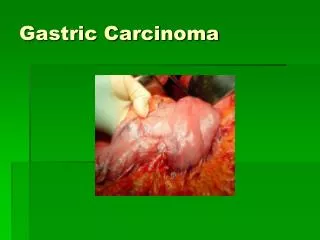
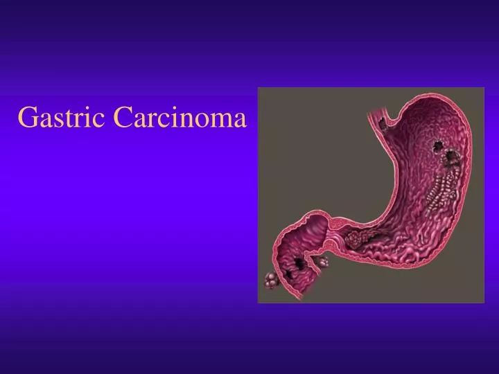
Gastric Carcinoma. Background. Second most common cancer-related death. Korea, Japan, China, Taiwan high rates. 22,000 diagnosed annually in US. 14 th most common cancer. Difficult to cure, as advanced disease. Most die of recurrent disease even after resection for cure. Anatomy.

E N D
Background • Second most common cancer-related death. • Korea, Japan, China, Taiwan high rates. • 22,000 diagnosed annually in US. • 14th most common cancer. • Difficult to cure, as advanced disease. • Most die of recurrent disease even after resection for cure.
Anatomy • Stomach begins at GE junction, ends at duodenum. • 3 parts- uppermost is cardia, largest part in middle is body, the last part is pylorus. • Cardia contains mucin producing cells. • Fundus or body mucoid cells, chief cells, parietal cells. • Pylorus has mucin producing cells.
Anatomy • Five layers: Mucosa, submucosa, muscular layer, subserosal layer, serosal layer. • Peritoneum of greater sac covers anterior surface • A portion of lesser sac drapes posteriorly over stomach. • The GE junction has limited serosal covering.
Anatomy • The site of the lesion is classified on basis of relationship to long axis of stomach. • 40% lower part • 40% middle part • 15% upper part • 10% more than one part • Recently the # of lesions proximally has increased.
Pathophysiology • Understand vascular supply, allows for understanding of routes of spread. • Derived from celiac artery. • Left gastric supplies upper right stomach. • Right gastric off common hepatic- lower portion. • Right gastroepiploic -lower portion of greater curve.
Pathophysiology • Understanding lymphatic drainage can clarify nodal involvement. • Complex drainage • Primarily along celiac axis. • Minor drainage along splenic hilum, suprapancreatic nodal groups, porta hepatis, and gastroduodenal areas
Frequency • US: seventh leading cause of cancer deaths, with 22,000 diagnosed yearly, and 14,000 deaths. • Internationally: second most common cancer. Tremendous geographic variation, with highest death rates in Chile, Japan, and former USSR.
Mortality and Morbidity • 5-year survival for curative resections ranges from 30-50% for stage II disease and 10-25% in stage III. • High likelihood of systemic and local relapse. • Adjuvant therapy is offered . • Operative mortality is less than 3% for curative resections.
Race • Higher in Asian countries. • Japanese detect patients at very early stage, patients appear to do quite well. • In Asian studies, patients with resected stage II and III disease have better outcomes than similar stages in the west. • Some believe this reflects a biologic difference between diseases in Asia and west. • Black race, low socioeconomic class.
Sex, Age • Men>women • Most are elderly at diagnosis. Median age 65 years. The ones that present in younger patients may represent a more aggressive variant. • Cigarettes
History • Early disease has no symptoms, some patients with incidental complaints get an early diagnosis. • If symptoms, it reflects advanced disease; These may include indigestion, nausea, dysphagia, early satiety, anorexia, weight loss.
History • Late complications include: pleural effusions, peritoneal effusions, GOO, GE obstruction, SBO, bleeding, jaundice, cachexia.
Physical • All physical signs are late events. • Too late for curative procedures. • Palpable stomach with succussion splash, hepatomegaly, Virchow nodes, sister MJ nodes, Blumer shelf, weight loss, pallor from bleeding and anemia.
Etiology • Diet • H. Pylori • Previous stomach surgery • Pernicious anemia • Polyps(rarely a precursor) • Atrophic gastritis • Radiation, genetics
Diet • Certain diets are implicated. • Rich in pickled vegetables, salted fish, excessive dietary salt, smoked meats. • A diet that includes fruits and vegetables rich in vitamin C may have a protective effect.
Helicobacter • Implicated as precursor of gastric cancer. • H. Pylori associated with atrophic gastritis, and patients with a history of prolonged gastritis have a 6-fold increase in risk. • Particularly true of tumors of antrum, body, and fundus of stomach, but not in cardia.
Previous Surgery • Implicated as risk factor, the rational being that previous gastric surgery alters normal pH of stomach. • Retrospective studies show that a small percentage of patients who have a gastric polyp removed have evidence of invasive carcinoma in the polyp. • Polyps may therefore be premalignant.
Genetic Factors • Poorly understood • Some familial aggregation exists
Laboratory • Assists in determining optimal therapy. • CBC identifies anemia, with may be caused by bleeding, liver dysfunction, or poor nutrition. • 30% have anemia. • Electrolyte panels and LFTs are also essential to better characterize patients clinical state.
Imaging Studies • EGD: safe, simple, providing a permanent color photographic record. • Obtains tissue for diagnosis. • UGI: detects large tumors, but only occasionally detects extension into esophagus or duodenum, especially if small or submucosal.
Imaging Studies • CXR: done to evaluate for metastases. • CT scan or MRI of chest, abdomen, pelvis: evaluate local disease process, and areas of spread. Some tumors are deemed unresectable based on the testing. • Accurately predicts stage 66-77%. • Poor nodal status prediction.
Endoscopic Ultrasound • Endoscopic ultrasound: becoming extremely useful as a staging tool, when CT fails to show T3, T4, or metastatic disease. • Used with neoadjuvant chemo to stratify pts • Can achieve resolution of 0.1 mm. • Cannot reliably distinguish between tumor and fibrosis. • Overall staging accuracy of 75% • Poor for T2 lesions (38%) • Better for T1(80%), T3 (90%)
Histology • Adenocarcinoma 95% • Lymphomas 2% • Carcinoids 1% • Adenocathomas 1% • Squamous cell 1%
Histology • Adenocarcinoma is classified according to the most unfavorable microscopic element present: tubular, papillary, mucinous, signet-ring cells. • Also identified by gross appearance: ulcerative, polypoid, scirrous, superficial spreading, multicentric, or Barrett ectopic. • Variety of other schemes: Borrmann, Lauren.
Borrmann Classification • 5 categories • Type I: polypoid or fungating • Type II: ulcerating lesions with elevated borders • Type III: ulceration with invasion of wall • Type IV: diffuse infiltration • Type V: cannot be classified
Lauren System • Epidemic or endemic • The intestinal, expansive epidemic type gastric cancer is associated with atrophic gastritis, retained glandular structure, little invasiveness, sharp margins. It would be a Borrmann I or II.
Lauren System • The epidemic or Borrmann I or II carries better prognosis, shows no family history. • The diffuse, infiltrative, endemic, is poorly differentiated, with dangerously deceptive margins, invades large areas of stomach. Younger patients, genetic factors, blood groups, and family history.
Staging • Primary tumor Tx- cannot be assessed T0- no evidence Tis- carcinoma in situ, no invasion of lamina T1- invades lamina propria or submucosa T2- invades muscularis or subserosa T3- penetrates serosa, no adjacent structure T4- invades adjacent structures
Regional Lymph Nodes NX- cannot be assessed N0- no nodes N1- mets in 1-6 regional nodes N2- mets in 7-15 regional nodes N3- mets in more than 15 regional nodes
Distant Metastases • MX- cannot be assessed • M0- no distant metastases • M1-distant metastases
Prognostic Features • Depth of invasion through gastric wall, presence or absence of regional lymph node involvement • The greater number of positive nodes, the greater the likelihood of local or systemic failure postoperatively
Spread Patterns • Directly, via lymphatics, or hematogenously • Direct extension into omentum, pancreas, diaphragm, transverse colon, and duodenum. • If lesion extends beyond wall to a free peritoneal surface, peritoneal involvement is frequent.
Spread Patterns • The visible gross lesion frequently underestimates true extent. • Abundant lymphatic channels in submucosal and subserosal layers allow for easy spread. • The submucosal plexus is prominent in esophagus, the subserosal plexus prominent in duodenum, which allows for proximal and distal spread. • Liver mets common, from hematogenous spread.
Laparoscopy • Inspect peritoneal surfaces, liver surface. • Identification of advanced disease avoids non-therapeutic laparotomy in 25%. • Patients with small volume metastases in peritoneum or liver have a life expectancy of 3-9 months, thus rarely benefit from palliative resection.
Lymph Node Dissection • AJCC: number rather than location of LN is prognostic. • Extent of dissection controversial. • Nodal involvement indicates poor prognosis, and more aggressive approaches to remove them are taking favor. • Ongoing trials regarding this in Europe. • Critics argue that the apparent benefit associated with extended LND reflects stage migration (each LN is reviewed more carefully).
Residual Disease R Status • Tumor status following resection. • Assigned based on pathology of margins. • R0- no residual gross or microscopic disease. • R1- microscopic disease only. • R2- gross residual disease. • Long term survival only in R0 resection.
“D” Nomenclature • Describes extent of resection and lymphadenectomy. • D1- removes all nodes within 3cm of tumor. • D2- D1 plus hepatic, splenic, celiac, and left gastric nodes. • D3- D2 plus omentectomy, splenectomy, distal pancreatectomy, clearance of porta hepatis nodes. • Current standards include a D1 dissection only.
Type of Surgery • In general most surgeons perform total gastrectomy ( if required for negative margins), esophagogastrectomy for tumors of the cardia and GE junction, and a subtotal gastrectomy for tumors of the distal stomach. • Similar 5 year rates for subtotal vs. total in tumors of distal stomach. • Extensive lymphatics require 5cm margin.
Outcome • 5-year survival for a curative resection is 30-50% for stage II disease, 10-25% for stage III disease. • Adjuvant therapy because of high incidence of local and systemic failure. • A recent Intergroup 0116 randomized study offers evidence of a survival benefit associated with postoperative chemoradiotherapy
Complications • Mortality 1-2% • Anastamotic leak, bleeding, ileus, transit failure, cholecystitis, pancreatitis, pulmonary infections, and thromboembolism. • Late complications include dumping syndrome, vitamin B-12 deficiency, reflux esophagitis, osteoporosis.
Adjuvant Therapy • Rationale is to provide additional loco-regional control. • Radiotherapy- studies show improved survival, lower rates of local recurrence when compared to surgery alone. • In unresectable patients, higher 4 year survival with mutimodal tx, in comparison to chemo alone.
Chemotherapy • Numerous randomized clinical trials comparing combination chemotherapy in the adjuvant setting to surgery alone did not demonstrate a consistent survival benefit. • The most widely used regimen is 5-FU, doxorubicin, and mitomycin-c. The addition of leukovorin did not increase response rates.