Download
1 / 26
270 likes | 494 Views
IMAGING OF MIDLINE TUMORS OF THE CENTRAL NERVOUS SYSTEM. F.Z. Belhoussine 1 , M. Boubbou 1 , B. Alami 1 , K.Moumna 2 , A.Amarti 2 , M.Benzegmout 3 , S. Tizniti 1. Department of Radiology 1, CHU Hassan II, Fez. Department of anatomo-pathology2 CHU Hassan II, Fez.

E N D
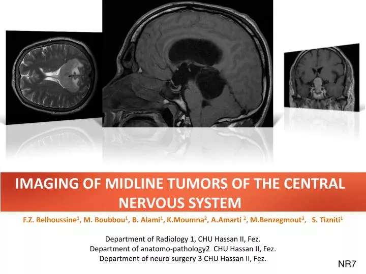
IMAGING OF MIDLINE TUMORS OF THE CENTRAL NERVOUS SYSTEM F.Z. Belhoussine1, M. Boubbou1, B. Alami1, K.Moumna2, A.Amarti2, M.Benzegmout3, S. Tizniti1 Department of Radiology 1, CHU Hassan II, Fez. Department of anatomo-pathology2 CHU Hassan II, Fez. Department of neuro surgery 3 CHU Hassan II, Fez. NR7
INTRODUCTION: • The anatomy of the supratentorielmidline structures of the brainiscomplex: • Corpus callosum, • Thirdventricle, • Trigone, • Pituitary gland • Pineal gland,… • Different types of tumorscan arise fromthese structures includingtumors of the trigone and septum, tumors of the falx, thirdventriculartumors and pinalregiontumors. • Thesetumorssharesimilarfeatures: minimal clinicalsymptomsdespitetheiroccasional large size, mild non-specificintracranial hypertension syndrome, value of MRI for depiction of tumor location, stereotacticbiopsy, relative difficulty of surgical management. 1 of 25 IMAGING OF MIDLINE TUMORS OF THE CENTRAL NERVOUS SYSTEM
OBJECTIVES: Illustrate the radiological semiology encephalic tumors of the midline. Show the value of MRI in the exploration of these tumors. 2 of 25 IMAGING OF MIDLINE TUMORS OF THE CENTRAL NERVOUS SYSTEM
BACKGROUND: • The anatomy of the supra tentoriel midline structures of the brain is complex. • We can distinguish 4 region: • Sellar and suprasellar region. • Corpus callosum region. • Intraventricular region. • Pineal region. • Falx region. • Brain CT is less sensitive to MRI to contribute for depiction of tumor location, stereotactic biopsy, relative difficulty of surgical management. 3 of 25 IMAGING OF MIDLINE TUMORS OF THE CENTRAL NERVOUS SYSTEM
BACKGROUND: ANATOMY : 5 2 1 4 3 B A Fig A: Coronal Tl MR ; 1: V3 region , 2:corpus callosum region , 5:falx region Fig B: Sagittal Tl MR ; 4: pineal region , 3: sellar and suprasellar region 4 of 25 IMAGING OF MIDLINE TUMORS OF THE CENTRAL NERVOUS SYSTEM
BACKGROUND: THE MAIN ETIOLOGIES by REGIONS : 5 of 25 IMAGING OF MIDLINE TUMORS OF THE CENTRAL NERVOUS SYSTEM
BACKGROUND: THE MAIN ETIOLOGIES by REGIONS : 6 of 25 IMAGING OF MIDLINE TUMORS OF THE CENTRAL NERVOUS SYSTEM
MATERIALS AND METHODS : A retrospective study involving 36 patients collected for department of radiology and neuro surgery over 3 years. Median age is 30 years (2- 77 years). In all patients conventional magnetic resonance imaging in conjunction with diffusion-weighted imaging (DWI) was performed. Proton magnetic resonance spectroscopy (MRS) was added in most cases. MRI examinations were performed in a single session on a machine GE 1.5 Tesla. Conventional MRI included: sagittal T1, coronal T2, axial FLAIR, axial T2 *, Diffusion, 3 planes T1 gadolinium and sequencespectroscopic. Histological confirmation was performed in all patients. 7 of 25 IMAGING OF MIDLINE TUMORS OF THE CENTRAL NERVOUS SYSTEM
RESULTS : 8 of 25 IMAGING OF MIDLINE TUMORS OF THE CENTRAL NERVOUS SYSTEM
RESULTS : Sellar /Suprasellarregion SUPRASELLAR MASS ADULT MRI sagittal section (A) and coronal (D) T2 W1 and coronal T1 without (B) and with gadolinium (C) showing a sellar and suprasellar mass isointense T1, T2 discrete, soisraisinghomogeneousaftercontrast. Selecteddiagnosis: pituitarymacroadenoma. Coronal graphic shows pituitarymacroadenoma (open arrow). Indentation from diaphragma sella causes "snowman" appearance (curvedarrows). Somecysticdegeneration & hemorrhageisdepicted. A B D C Anapath : PITUITARY MACROADENOMA 09 of 25 IMAGING OF MIDLINE TUMORS OF THE CENTRAL NERVOUS SYSTEM
RESULTS : SUPRASELLAR MASS CHILD Sellar /Suprasellarregion MRI in coronal (A) and sagittal (D) T1, coronal T2 (B) and T1 contrastCornale (c) objectifying the presence of a large sellar and suprasellarprocesswith a heterogeneousisointense T1 (A and D), discretehyperintense T2 (B), withenhancement of heterogeneouslyaftercontrast (C). thisprocesspresents an important extension latérosellaireresponsiblehydrocephalusupstream. Sagittal graphic shows a predominantly cystic, partially solid, suprasellar mass with focal rim calcifications. Note small intrasellar component and fluid-fluid level. A B C D Anapath : CRANIOPHARYNGIOMA 10 of 25 IMAGING OF MIDLINE TUMORS OF THE CENTRAL NERVOUS SYSTEM
RESULTS : Sellar /Suprasellarregion CRANIOPHARYNGIOMA • Benign dysontogenetic epithelial tumor derived from Rathke pouchepithelium. • Two types: Adamantinomatous and papillary. • Location: • Surgical division of craniopharyngioma into three groups: Sellar/ Prechiasmatic / Retrochiasmatic. • Best diagnostic clue: • CT Finding: Partially Ca++, partially solid, cystic • suprasellar mass in a child. • MR Finding: • - High signal intensity suprasellar mass on pre-contrast T1WI • - Tl C+: Solid portions enhance heterogeneously, cyst wallsenhancestrongly. 11 of 25 IMAGING OF MIDLINE TUMORS OF THE CENTRAL NERVOUS SYSTEM
RESULTS : Sellar /Suprasellarregion CRANIOPHARYNGIOMA • Pathology: • Most common pediatric intracranial tumor of non-glial origin. • Approximately 54% of all pediatric sellar/chiasmaticregiontumors are CPs • Clinical Issues: • Clinical profile: Pediatric patient with morning headache, visual defect, short stature. • Age: Bimodal age distribution (peak 5-15 Years papillary CP > SOy). • 64-96% overall 10 year survival. • Diagnostic Checklist: • Use NECT to detect Ca++ if MR diagnosis is in question. 12 of 25 IMAGING OF MIDLINE TUMORS OF THE CENTRAL NERVOUS SYSTEM
RESULTS : Sellar /Suprasellarregion D A Fig A,B, C and D show a sphenoidridge mass slightly intense with cortex in axial and sagittal T1W1(fig A, D) and axial T2W1(Fig C)witchenhancehomogeneously & intensely (fig B) C Anapath:MENINGIOMA B 13 of 25 IMAGING OF MIDLINE TUMORS OF THE CENTRAL NERVOUS SYSTEM
RESULTS : Intraventricular (3rdventricle) T2 Axial graphic shows a classic CC at the foramen of Monro causingmild/moderate obstructive hydrocephalus. Note fornices and choroid plexus are elevated, stretched over the cyst (arrows). D A 31 years old. Headaches with visual disturbances neglected for a year. MRI: round lesion of the anterior third ventricle, although limited in homogeneous hyperintense T1 and T2, clogging the holes of Monro, causing a biventricular hydrocephalus active upstream (FLAIR hyper intense peri-ventricular). C Coronal T1 Flair W1 Anapath: COLLOID CYST 14 of 25 IMAGING OF MIDLINE TUMORS OF THE CENTRAL NERVOUS SYSTEM
RESULTS : Intraventricular (3rdventricle) D A Coronal T1W1 Gado+ Coronal T1 Flair W1 Axial FSPGR 3D C 26 yearsold, Headache, nausea and vomiting, MR find a mass in the thirdventricle, hypo intense of Gray matter in T1W1, hyper intense to CSF in Flair and ringlikeenhancement of cyst. Anapath:PILOCYTIC ASTROCYTOMA 15 of 25 IMAGING OF MIDLINE TUMORS OF THE CENTRAL NERVOUS SYSTEM
Intraventricular (3rdventricle) RESULTS : T2 38 years old. Headaches with visual disturbances and vomiting. MRI: IntraventricularWelldelineated, lobulated mass in thirdventricle iso- intense In T1W1, hyper intense in T2W1, strongly and homogeneouslyenhanced. D SAG t1 SAG T1 GADO + A • Typically pediatric tumors, lateral ventricle • In adults, 4th ventricle, thirdventricle in 10% • Enhancing papillary mass, hydrocephalus common C Coronal T1 Flair W1 Anapath: PAPILLOMA COR T2 flair COR T1 GADO + 16 of 25 IMAGING OF MIDLINE TUMORS OF THE CENTRAL NERVOUS SYSTEM
RESULTS : Ca+ Pinealregion T2 Axial CT •Engulfs" calcifiedpineal gland • Intensely enhancing pineal mass, often homogeneous • Often CSF spread at diagnosis • Hyperdense on CT • Typicallyyoung male patients D SAG T1 SAG T1 Gado+ A SAG T1 GADO + SAG t1 14 years old. Headaches with visual disturbances and vomiting. MRI: pineal region mass arround the posterior third ventricle with calcifications in CT, iso- intense In T1W1, hyper intense in T2W1, Slightlyhyperintense flair , causing a triventricular hydrocephalus active upstream and homogeneouslyenhanced. Axial T2 flair C Anapath: Germinoma COR T2 17 of 25 IMAGING OF MIDLINE TUMORS OF THE CENTRAL NERVOUS SYSTEM
RESULTS : Pinealregion •Round, smoothcystic mass • Typically < 1 cm, may be up to 2 cm • Variable calcification and cyst fluid • No or minimal rimenhancement, compressedenhancing gland often seen posteriorly • May be indistinguishable from PC on imaging T2 D A SAG T1 GADO + SAG t1 SAG T1 AXIAL T2 COR T1 31 years old. Fainting, convulsive crisis last year. MRI: Round cyst hypo intense T1, hyper intense T2 without calcifications. C PINEAL CYST 18 of 25 IMAGING OF MIDLINE TUMORS OF THE CENTRAL NERVOUS SYSTEM
RESULTS : Pinealregion • Most epidermoid cysts resemble CSF, not fat • No dermalappendages • 4-9x more common than dermoid • Off-midline> midline: 40-50% in CPA, 10-15% para sellar/middle fossa, 10% diploic • MRI: Isointense to CSF exceptrestricts on diffusion T2 Axial T2 D COR T1 COR flair T2 A SAG T1 GADO + SAG t1 37 years old. Headache. MRI: lobular mass occupatingtrijimenal region slightlyhyperintense to CSF on T1W1, doesn’t completely null on flair causing triventricular hydrocephalus active upstream, hyper intense on T2 , without enhancement. v4 SAGT1 gado + C Anapath: EPIDERMOID CYST 19 of 25 IMAGING OF MIDLINE TUMORS OF THE CENTRAL NERVOUS SYSTEM
RESULTS : Pinealregion SAG T1 Cor T1 gado+ Cor T2 flair 14 years old. Headache. .MRI: lobular mass occupatingtrijimenal region slightlyhyperintense to CSF on T1W1 flair, causing triventricular hydrocephalus active upstream, hyper intense on T2 flair, without enhancement. C Coronal T1 Flair W1 Anapath: ASTROCYTOMA LOW GRADE Axial SPGR 20 of 25 IMAGING OF MIDLINE TUMORS OF THE CENTRAL NERVOUS SYSTEM
RESULTS : Trigone and corpus callosum region • •Periventricular enhancing mass • •Often crosses corpus callosum • •Typically iso-intense/hypointense on T2WI • • Necrosis common in AIDS related lymphoma. • Intravascular lymphoma may appear diffusely infiltrating. T2 Axial T2 D COR T1 COR flair T2 A SAG T1 GADO + SAG t1 Axial flair T1 Axial T2 Axial T1 Gado + v4 55 years old. Headache and memory troubles. MRI: enhancing lesion involving the entire corpus callosum showing minimal mass effect on the ventricle. Contrast enhanced axial T1W1. C Coronal T1 Flair W1 Anapath: LYMPHOMA 21 of 25 IMAGING OF MIDLINE TUMORS OF THE CENTRAL NERVOUS SYSTEM
RESULTS : Trigone and corpus callosum region T2 Axial T2 D COR T1 A SAG T1 GADO + SAG t1 Sagittal T1 Coronal T1 Flair W1 TDM axial C+ 55 years old. Headache and memory troubles. MRI: Heterogenous mass involving the entire corpus callosum . It’s hypo intense on T1W1 and shows heteregenous enhancement lesion involving the entire corpus callosum showing minimal mass effect on the ventricle. Contrast enhanced axial T1W1. C Anapath: CORPUS CALLOSUMGLIOMA 22 of 25 IMAGING OF MIDLINE TUMORS OF THE CENTRAL NERVOUS SYSTEM
CONCLUSION : Tumors of the midline of the brain are complex and diverse. The magnetic resonance imaging is of major interest, allowing a particularly informative topographic analysis, a diagnostic aid, bringing sometimes a tissue characterization and support the choice of treatment, specifying the path and the target in stereotactic biopsies. COR T1 A SAG t1 23 of 25 IMAGING OF MIDLINE TUMORS OF THE CENTRAL NERVOUS SYSTEM
BIBLIOGRAPHY : 1- A.Osborn, S.Blaser, K.Salzman. Diagnostic Imaging :Brain. 2- Tumeurscranioencéphalique de la lignemédiane. C.Delmaire; JY.Gauvrit; EL Hajj; G.Soto.Ares; JR2006, 87: 764- 78. 3- Tous en selle sur l’hypophyse. R.Richard, C. Vandendries, F. Benoudiba, N. Hocine, M. Adoui, G. Nasser, D. Ducreux. SFR 2011. 4- Imagerie des tumeurs de la région pinéale : à propos de 26 cas S. Belkacem, M. Fikri, J. FaikOuahab, N. Ech-Cherif El Kettani, MR. El Hassani, M. Jiddane . SFR2011 . 5- Aide au diagnostic des lésions kystiques intracrâniennes .Y Alaoui Lamrani, M Maâroufi, I Kamaoui, N Hammas, H Ammor, L Benjelloun, M Boubbou, N Sqalli Houssaini, A Amarti, S Tizniti. SFR 2011. COR T1 A SAG t1 24 of 25 IMAGING OF MIDLINE TUMORS OF THE CENTRAL NERVOUS SYSTEM
THANKS COR T1 A SAG t1 25 of 25 IMAGING OF MIDLINE TUMORS OF THE CENTRAL NERVOUS SYSTEM