Download
1 / 36
430 likes | 1.52k Views
Chapter 15 - Genetics of Bacteria and Bacteriophages : Mapping bacteria, 3 different methods : Conjugation Transformation Transduction Bacteriophage mapping : Bacteriophage gene mapping Cis -trans complementation test. Bacteria transfer (or receive) genetic material 3 different ways :

E N D
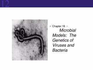
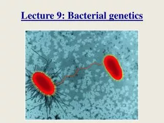
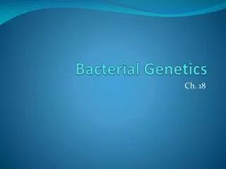
Chapter 15 - Genetics of Bacteria and Bacteriophages: Mapping bacteria, 3 different methods: • Conjugation • Transformation • Transduction Bacteriophage mapping: • Bacteriophage gene mapping • Cis-trans complementation test
Bacteria transfer (or receive) genetic material 3 different ways: • Conjugation • Transformation • Transduction • Transfer of DNA always is unidirectional, and no complete diploid stage forms.
Conjugation: Discovered by Joshua Lederberg and Edward Tatum in 1946. Unidirectional transfer of genetic material between donor and recipient bacteria cells by direct contact. Segment (rarely all) of the donor’s chromosome recombines with the homologous recipient chromosome. Recipients containing donor DNA are called transconjugants.
Fig. 15.2, Lederberg & Tatum (1946) Experiment demonstrating recombination in E. coli. Recombination of 2 complimentary auxotrophs gives rise to a strain that can synthesize all nutrients.
Fig. 15.3, Bernard Davis experiment demonstrated that physical contact is required for bacterial recombination.
Conjugation-transfer of the sex factor F: William Hayes (1953) demonstrated that genetic exchange in E. coli occurs in only one direction. Genetic transfer is mediated by sex factor F. Donor is F+ and recipient is F-. F is a self-replicating, circular DNA plasmid (1/40 the size of the main chromosome). F plasmid contains an origin sequence (O), which initiates DNA transfer. It also contains genes for hair-like cell surface (F-pili or sex-pili), which aid in contact between cells. No conjugation can occur between cells of the same mating type. Conjugation begins when the F plasmid is nicked at the origin, and a single strand is transferred using the rolling circle mechanism. When transfer is complete, both cells are F+ double-stranded.
Figs. 15.4 & 15.5a Transfer of the F factor
Conjugation of high-frequency recombinant strains: No chromosomal DNA is transferred by standard sex factor F. Transfer of chromosome DNA is facilitated by special strains of F+ integrated into the bacteria chromosome by crossing over. Hfr strains = high frequency recombination strains. Discovered by William Hayes and Luca Cavalli-Sforza. Hfr strains replicate F factor as part of their main chromosome. Conjugation in Hfr strains begins when F+ is nicked at the origin, and F+ and bacteria chromosomal DNA are transferred using the rolling circle mechanism. Complete F+ sequence (or complete chromosomal DNA) is rarely transferred (1/10,000) because bacteria separate randomly before DNA synthesis completes. Recombinants are produced by crossover of the recipient chromosome and donor DNA containing F+.
Fig. 15.5b Transfer of the HfrF+ factor
Fig. 15.6 Excision of the F+ factor also occurs spontaneously at low frequency. Begin with Hfr cell containing F+. Small section of host chromosome also may be excised, creating an F’ plasmid. F’plasmid is named for the gene it carries, e.g., F’ (lac)
Using conjugation to map bacterial genes: • Begin with two different Hfr strains selected from F+ x F-crosses and perform an interrupted mating experiment. • HfrH thr+ leu+ aziR tonR lac+ gal+ strR F-thr leu aziS tons lac gal strS • Mix 2 cell types in medium at 37°C. • Remove at experimental time points and agitate to separate conjugating pairs. • Analyze recombinants with selective media. • Order in which genes are transferred reflects linear sequence on chromosomes and time in media. • Frequency of recombinants declines as donor gene enters recipient later.
Fig. 15.7 Interrupted mating experiment
Fig. 15.7c, Genetic map-results of interrupted E. coli mating experiment.
Generating a map for all of E. coli: Location and orientation of the HfrF+ in the circular chromosome varies from strain to strain. Overlap in transfer maps from different strains allow generation of a complete chromosomal map. Fig. 15.8
Circular genetic map of E. coli Total map units = 100 minutes Time required for E. coli chromosome to replicate at 37°C.
Transformation: Unidirectional transfer of extracellular DNA into cells, resulting in a phenotypic change in the recipient. First discovered by Frederick Griffith (1928). DNA from a donor bacteria is extracted and purified, broken into fragments, and added to a recipient strain. Donor and recipient have different phenotypes and genotypes. If recombination occurs, new recombinant phenotypes appear.
More about transformation: Bacteria vary in their ability to take up DNA. Bacteria such as Bacillus subtilis take up DNA naturally. Other strains are engineered (i.e., competent cells). Competent cells are electroporated or treated chemically to induce E. coli to take up extracellular DNA.
Fig. 15.9, Transformation of Bacillus subtilus Heteroduplex DNA
Mapping using transformation: • Recombination frequencies are used to infer gene order. p+ q+ o+ x p q o • If p+ and q+ frequently cotransform, order is p-q-o. • If p+ and o+ frequently cotransform, order is p-o-q.
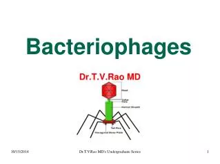
Transduction: • Bacteriophages (bacterial viruses) transfer genes to bacteria (e.g., T2, T4, T5, T6, T7, and ). • Generalized transduction transfers any gene. • Specialized transduction transfers specific genes. • Phages typically carry small amounts of DNA, ~1% of the host chromosome. • Viral DNA undergoes recombination with homologous host chromosome DNA.
Fig. 15.12 Life cycle of phage
Fig. 15.13 Generalized transduction of E. coli by phage P1
Transduction mapping is similar to transformation mapping: • Gene order is determined by frequency of recombinants. • If recombination rate is high, genes are far apart. • If recombination rate is low, genes are close together.
Mapping genes of bacteriophages (see Fig. 15.15): Infect bacteria with phages of different genotypes using two-, three-, or four-gene crosses crossover. Count recombinant phage phenotypes by determining differences in cleared areas (no bacteria growth) on a bacterial lawn. Different phage genes induce different types of clearing (small/large clearings with fuzzy/distinct borders).
Fine structure gene-mapping of bacteriophages: Same principles of intergenic mapping also can be used to map mutation sites within the same gene, intragenic mapping. First evidence that the gene is sub-divisible came from C. P. Oliver ‘s (~1940) work on Drosophila. Seymour Benzer’s (1950-60s) study of the rII region of bacteriophage T4.
Seymour Benzer’s (1950-60s) study of the rII region of T4: • Studied 60 independently isolated rII mutants crossed in all possible combinations. • Began with two types of traits: plaque morphology and host range property. • Growth in permissive host E. coli B; all four phage types grow. • Growth in non-permissive hostE. coli K12();rare r+ recombinants grow (rare because the mutations are close to each other and crossover is infrequent). • Benzer studied 3000 rII mutants showing nucleotide deletions at different levels of subdivision (nested analyses). • Was able to map to T4 to level equivalent to 3 bp (the codon). • Ultimately determined that the rII region is sub-divisible into >300 mutable sites by series of nested analyses and comparisons.
Benzer identified recombinants of two rII mutants of T4 using different strains of E. coli.
Fig. 15.18, Benzer’s map of the rII region generated from crosses of 60 different mutant T4 strains.
Fig. 15.19 Benzer’s deletion analysis of the rII region of T4: No recombinants can be produced if mutant strain lacks the region containing the mutation.
Fig. 18.20 (2nd edition), Benzer’s deletion map divided the rII region into 47 segments.
Fig. 15.20, Benzer’s composite map of the rII region indicating >300 mutable sites on two different genes. Small squares indicate point mutations mapping to a given site.
Seymour Benzer’s cis-trans complementation test: Used to determine the number of functional units (genes) defined by a given set of mutations, and whether two mutations occur on the same unit or different units. If two mutants carrying a mutation of different genes combine to create a wild type function, two mutations compliment. If two mutants carrying a mutation of the same gene create a mutant phenotype, mutations do not compliment.
Fig. 15.21, Seymour Benzer’s cis-trans complementation test.