Download
1 / 34
370 likes | 711 Views
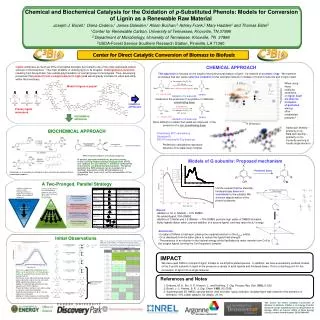
Monomers, polymers, and macromolecules. There are 4 categories of macromolecules: Carbohydrates Proteins, Lipids, and Nucleic acids. Carbon is the central element. All biomolecules contain a Carbon chain or ring Carbon has 4 outer shell electrons (valence = 4)

E N D
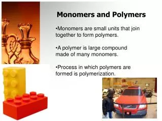
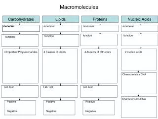
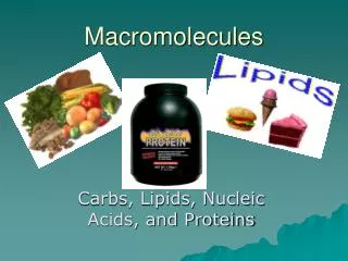
Monomers, polymers, and macromolecules There are 4 categories of macromolecules: Carbohydrates Proteins, Lipids, and Nucleic acids
Carbon is the central element • All biomolecules contain a Carbon chain or ring • Carbon has 4 outer shell electrons (valence = 4) • Therefore it’s bonding capacity is great • It forms covalent bonds –hence, has strong bonds • Once bound to other elements (or to other Carbons), it is very stable
Carbon linkages CH4 = Propane • Single chains • Rings = C3H8 The 4 types of biomolecules oftenconsist of large carbon chains
Carbon binds to more than just hydrogen!! • To OH groups in sugars • To NH2 groups in amino acids • To H2PO4 groups of nucleotides of DNA, RNA, and ATP Amino acid OH, NH2, PO4 are called ‘functional groups’!
Fig. 3.1 Functional groups:
Isomers have the same molecular formulas but different structures • Structural isomer = difference in the C skeleton structure • Stereoisomer = difference in location of functional groups
Enantiomers are special types of stereoisomers Enantiomers are mirror images of each other One such enantiomer contains C bound to 4 different molecules and is called a chiral molecule Chiral molecules rotate polarized light to the right (D form) or to the left (L form) molecules Examples: amino acids (L form) sugars (D form)
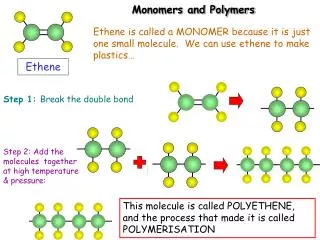
Monomers and polymers • Monomers are made into polymers via dehydration reactions • Polymers are broken down into monomers via hydrolysis reactions
Carbohydrates (or sugars) • Simple sugars (monosaccharides) • Only one 3-C, 5-C, 6-C chain or ring involved
Fig. 3.5 Examples of sugar monomers* *Remember how C’s are counted within the ring structures (starting from the right side and counting clockwise)
Carbohydrates (sugars) • Double sugars (disaccharides) • Two 6-C chains or rings bonded together
Carbohydrates (sugars) • Complex carbo’s (polysaccharides) • Starch • Cellulose • Glycogen • Chitin Glycogen to glucose in animals
Fig. 3.9 Polysaccharides Starch structure vs Glycogen structure
Fig. 3.10 Polysaccharides: Cellulose structure
Proteins • Composed of chains of amino acids • 20 amino acids exist • Amino acids contain • Central Carbon • Amine group • Carboxyl group • R group
Fig. 3.20 The 20 Amino Acids All differ with respect to their R group
Peptide bonds occur between amino acids • The COOH group of 1 amino acid binds to the NH2 group of another amino acid • Forms a peptide bond!
Fig. 3.21 The chain (polymer) of amino acids forms a variety of loops, coils, and folded sheets from an assortment of bonds and attractions between amino acids within the chain(s)
There are at least 7 functions of proteins • Enzyme catalysts – specific for 1 reaction • Defense – antibody proteins, other proteins • Transport- Hgb, Mgb, transferrins, etc • Support – keratin, fibrin, collagen • Motion – actin/myosin, cytoskeletal fibers • Regulation- some hormones, regulatory proteins on DNA, cell receptors • Storage – Ca and Fe attached to storage proteins
There are four levels of protein structure • Primary = sequence of aa’s • Secondary = forms pleated sheet, helix, or coil • Tertiary = entire length of aa’s folded into a shape • Quaternary = several aa sequences linked together
Fig. 3.23 Motifs and Domains: Important features of 2° and 4° structure
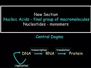
Nucleic acids: DNA and RNA • DNA = deoxyribonucleic acid • DNA is a double polymer (chain) • Each chain is made of nucleotides • The 2 chains bond together to form a helix
DNA nucleotides • Each nucleotide in DNA contains: • 5-C sugar (deoxyribose) • Phosphate • Nitrogen base -adenine (A) -guanine (G) -cytosine (C) -thymine (T)
Fig. 3.14 One polymer of nucleotides on one “backbone” of nucleic acid
Fig. 3.15 The DNA “double helix”
Lipids: Hydrophobic molecules • Central core of glycerol • Bound to up to 3 fatty acid chains • They exhibit a high number of C-H bonds – therefore much energy and non-polar • When placed in water, lipids spontaneously cluster together • They help organize the interior content of cells “phospholipids”
Glycerol and fatty acid chains What specific bonds form between glycerol and each fatty acid chain? Would you think this to be an hydrolysis or a dehydration synthesis rxn?
Saturated and unsaturated fats The difference resides in the number of H’s attached to C’s in the fatty acid chains; the amount of “saturation” on the C’s
Saturated vs unsaturated fats and diet • Saturated fats raise LDL-cholesterol levels in the blood (animal fats, dairy, coconut oil, cocoa butter) • Polyunsaturated fats leave LDL-cholesterol unchanged; but lower HDL-cholesterol (safflower and corn oil) • Monounsaturated fats leave LDL and HDL levels unchanged (olive oil, canola, peanut oil, avocados) • One variety of polyunsaturated fat (Omega-3 fatty acids) guards against blood clot formation and reduce fat levels in the blood (certain fish, walnuts, almonds, and tofu)
Phospholipids and cell membranes • P-lipids make up the majority of cell membranes including: • The plasma membrane • Nuclear envelope • Endoplasmic reticulum (ER) • Golgi apparatus • Membrane-bound vesicles
Structure of single P-lipid The 3 C’s of glycerol are bound to: 2 fatty acid chains phosphate
Cell environment organizes P-lipid bilayer to proper orientation Hydrophilic (polar) “heads” of P-lipid oriented to the exterior; hydrophobic (non-polar) “tails” oriented to the interior