Download
1 / 41
520 likes | 2.34k Views
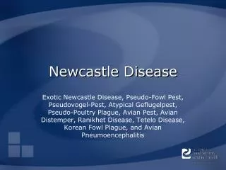
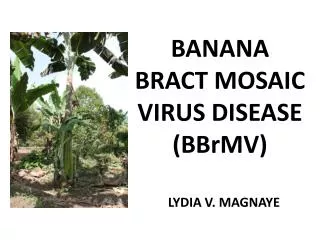
NEWCASTLE DISEASE VIRUS. Presented To: Dr. Madiha Salah Ebrahim. Edit by: 1. Mai Labib Badr. 6. Nehad Ahmed Saleh. 2. Nesma Khatan. 7. Maha Tantawy. 3. Maha El.Ansary. 8. Amina El.Nagar.

E N D
NEWCASTLE DISEASE VIRUS Presented To: Dr. Madiha Salah Ebrahim. Edit by: 1. Mai Labib Badr. 6. Nehad Ahmed Saleh. 2. Nesma Khatan. 7. Maha Tantawy. 3. Maha El.Ansary. 8. Amina El.Nagar. 4. Amira Abd El.Naser. 9. Hadeer Fouad Shams. 5. Lamiaa Gamal. 10. Amal Samy.
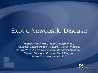
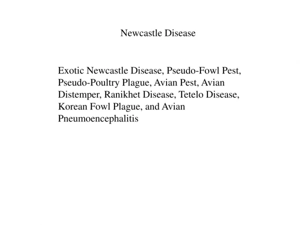
Newcastle disease virus • NDV also called (avian paramyxovirus type I, pneumoencephalitis virus & pseudo-fowl pest). • ND is contagious and fatal viral disease affecting most species of birds (chickens, turkeys, pigeons ,parrots ,ducks, geese, quails)and human. Taxonomy of the NDV : • Family: Paramyxoviridae. • Subfamily: Paramyxovirinae. • Genus: Avulavirus.
Characteristics: • Spherical virus with diameter of 100-300 nm. • Enveloped virus (containing lipid, CHO & protein). • It has segmented & single stranded negative sense RNA. • Two specific virus proteins (hemagglutinin-neuraminidase & fusion protein) are the main proteins found in the outer coat of the virus. • Replication occurs in the cytoplasm of the host cell. • Affected species; birds & human. • Morbidity; Up to100% & Mortality; 90%.
Fig1; Diagrammatic representation of Newcastle disease virus.
Inactivation of the virus: • Minimum core temperature of 80°C for one minute, 75°C for 5 minutes or 70°C for 30 minutes - completely destroys the virus in meat. • Ether sensitive and inactivated by formalin, phenol & acid pH. • Destroyed rapidly by dehydration and ultraviolet rays. CPE of NDV: • Syncytia formation. • Production of inclusion bodies. • Haemadsorption of G.pig RBCs by infected cell culture.
Haemagglutination: • All strains of NDV agglutinate (Chicken, G.pig, human group O)RBCs. • Most strains agglutinate (OX & sheep)RBCs. • Horse RBCs agglutinated by lentogenic strain. • NDV cause haemadsorption. Antigenic properties: • NDV is immunologically distinct from orthomyxoviruses & from other members of paramyxoviruses. • Mumps may develop HI antibodies to NDV.
Cultivation: • NDV is inoculated into 10-12 days hen embryonated eggs via chorioallantoic membrane or allantoic sac. • It produces haemorrhagic lesions and encephalitis & embryo dies within 34-72 hours. • NDV grows well in chicken embryo fibroblast cell culture. • Maximum titer is obtained after 24-36 hours. • Titer of the virus in tissue culture is one log lower than that in embryonated egg.
Strains of NDV classified according to their pathogenicity into: 1. Viscerotropic velogenic NDV (Doyle's form). 2. Neurotropic velogenic NDV (Beache's form). 3. Mesogenic NDV (Beaudett's form). 4. Lentogenic NDV (Hitchner form). 5. Asymptomatic enteric NDV.
Transmission: • Direct contact between healthy birds and the infected bird discharges. • Contaminated feed, water, equipment and clothing. • Virus can be picked up on shoes and clothing and carried from an infected flock to a healthy one. • Airborne spread. • Contaminated poultry vaccines. • Other animals and birds transporting the virus from farm to farm.
Pathogenicity: • the virulence of NDV Can be determined by the following techniques: 1. The mean death time in chicken egg embryo (MDT). 2. Intracerebral Pathogenicity index (ICPI) in day-old chick. 3. Intravenous Pathogenicity index (IVPI) in 6 week old chickens. 4. Intracloacal Pathogenicity test in 6-8 week old chickens.
Incubation period: • It varies from (2 to 15) days in poultry depending on the virulence of the strain. • In chickens infected with velogenic isolates; (2 to 6) days. • In some avian species; 25 days. Pathogenesis: • The virus replicates in the mucosa of the upper respiratory and intestinal tracts. • Virus spreads via blood to spleen and bone marrow (viremia) causing infection of other organs: lung ,intestines & C.N.S.
Immune response against NDV: • Antibody production is rapid. • HI antibody can be detected within 4-6 days of infection. • The level of HI antibody is a measure of immunity. • Serum antibodies of the hen are transferred to chicks through yolk, and protect chicks for 3-4 weeks after hatching. • Serum IgG does not prevent respiratory infection. • Locally produced IgA prevent respiratory and the intestinal infection.
Clinical Signs and Symptoms: • Respiratory symptoms. • Nervous signs. • Digestive symptoms. • Drop in egg production with thin, rough-shelled eggs. • Swelling of tissues around eyes and in the neck. • Sudden death. • In human;(Mild conjunctivitis, influenza-like symptoms and laryngitis).
Fig7; The chicken stand on its hock joints, a sign of generalized depression.
Fig8; Diarrhea with green bile pigment and white urates. Fig9; Square appearance of the head due to bilateral facial edema.
Fig10; Swelling of the lower eyelid and conjunctivitis. Fig11; Reddening of periocular region and corneal edema.
PM and gross lesions: • Inflammation with Petechial hemorrhages on proventriculus mucosa. • Edematous, hemorrhagic, necrotic, and ulcerative areas on Peyer's patches, caecal tonsils. • Edematous, hemorrhagic, or degenerated ovaries.
Fig13; subconjunctival haemorrhages with external lesions. Fig14; Odema and hemorrhages in the conjunctiva and infraorbital sinus.
Fig15; Accumulation of mucus in the respiratory tract. Fig16; Mild haemorrhagic lesions in the mucosa of trachea.
Fig17; Congestion and haemorrhages in the pharynx and proximal trachea. Fig18; Sever thymus atrophy with extensive haemorrhages.
Fig19; Inflammation with pinpoint heamorrhagic lesions in the proventriculus mucosa.
Fig20; Necrosis of lymphoid tissue at the caecal tonsils. Fig21; Extensive haemorrahges and ulcers of caecal tonsils mucosa.
Fig22; Acute focal lymphoid necrosis in the duodenum. Fig23; Focal ulceration and haemorrahge in the small intestine.
Fig24; Haemorrhagic lesions along the entire length of the intestine. Fig25; Sever haemorrhages in the rectal mucosa.
Fig26; Ulcers with fibrin accumulation in the mucosa of the cloaca . Fig27; Pulmonary congestion and edema.
Fig28; The spleen is enlarged with numerous white lesions. Fig29; Extensive haemorrhages on the liver.
Fig30; Odema and haemorrahges within the mucosa of bursa of fabricius. Fig31;Sever acute haemorrahges and congestion in the ovarian follicles.
Diagnosis: • It includes: 1. Clinical signs and symptoms. 2. Lab tests include; • Serological tests:Haemagglutination inhibition test, Enzyme Linked Immunosorbant Assay (ELISA), PCR & Sequence technology. • Pathogenicity assessment: • Plaque test in chicken embryo fibroblast cultures. • Mean death time. • Intracerebral pathogenicity index. • Intravenous pathogenicity index.
Diagnostic Samples: • Samples from live birds: • Tracheal swabs. • Cloacal swabs. • Faecal swabs. • Serum. • Samples from dead birds: • Lung, kidneys, intestine, spleen, brain, liver, and heart tissues.
Differential diagnosis: • Pathogenic avian influenza. • Infectious laryngotracheitis. • Salmonellosis & Mycoplasmosis. • Vit. E and Selenium deficiency. • Avian encephalomyelitis. • Infectious bronchitis. • Fowl cholera. • Fowl pox. • Coryza.
Treatment: • There is no known treatment for Newcastle Disease. Prevention: • Quarantine & isolation of all newly purchased birds for at least 30 days. • Transportation of birds in new or disinfected containers. • Restrict personnel movement between new and old birds. • Disinfection of all surfaces and equipment. • Disposal of any destroyed birds and contaminated products. • Removal of insects and mice (vectors). • Control handling of bird carcasses, litter and manure.
Vaccination: • Vaccines are administrated at 2 to 4 weeks of age or at 1 day of age via conjunctival instillation. • Vaccine-induced immunity is short-lived (8–10) weeks. • Live vaccine. • Inactivated vaccine. • Newplex (a proprietary virus antibody complex vaccine).
REFERENCES: • http://www.fst.osu.edu/li/research.htm • http://www.spc.int/rahs/Manual/AVIAN/NEWCASTLE.htm • http://www.daff.gov.au/animal-plant-health/pests-diseases-weeds/animal/newcastle • http://www.cfsph.iastate.edu/DiseaseInfo/clinical-signs-photos.php?name=newcastle-disease • www.webconferences.com/.../ppt/Newcastle%20Disease%20Virus_Samal.pdf • www.fao.org/docrep/003/t0756e/T0756E08.htm • Clubb, S. Laws and Regulations Affecting Aviculture and the Pet Bird Industry. Altman, R; Clubb, S; Dorrestein, G; Quesenberry, K. (eds.). Avian Medicine and Surgery. W.B. Saunders. Philadelphia, PA; 1997. • Gallerstein, G; Acker, H. The Complete Bird Owner's Handbook. Simon & Schuster Macmillan. New York, New York; 1994. • Olsen, G; Orosz, S. Manual of Avian Medicine. Mosby, Inc. St. Louis, MO; 2000. • Rupley, A. Manual of Avian Practice. W.B. Saunders. Philadelphia, PA; 1997. • USDA Department of Agriculture. Veterinary Services. Exotic Newcastle Disease. May, 2001.