Download
1 / 77
870 likes | 1.29k Views
Chapter 5 Proteins: Their Primary Structure and Biological Function. Essential Question. What structural forms do polypeptide chains assume, how can the sequence of amino acids in a protein be determined, and what are the biological roles played by proteins?. Outline.

E N D
Chapter 5Proteins: Their Primary Structure and Biological Function
Essential Question • What structural forms do polypeptide chains assume, how can the sequence of amino acids in a protein be determined, and what are the biological roles played by proteins?
Outline • What is the fundamental structural pattern in proteins? • What architectural arrangements characterize protein structure? • How are proteins isolated and purified from cells? • How is the amino acid analysis of proteins performed? • How is the primary structure of a protein determined? • Can polypeptides be synthesized in the laboratory? • What is the nature of amino acid sequences? • Do proteins have chemical groups other than amino acids? • What are the many biological functions of proteins?
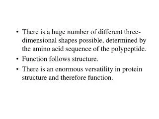
5.1 What Architectural Arrangements Characterize Protein Structure? • Proteins are classed according to shape and and solubility • Shape - globular or fibrous • The four levels of protein structure are: - Primary (1°) - sequence - Secondary (2°) - local structures - H-bonds - Tertiary (3°) - overall 3-dimensional shape - Quaternary (4°) - subunit organization
5.1 What Architectural Arrangements Characterize Protein Structure? (a) Proteins having structural roles in cells are typically fibrous and often water insoluble. (b) Myoglobin is a globular protein. (c) Membrane proteins fold so that hydrophobic amino acid side chains are exposed in their membrane-associated regions.
5.1 What Architectural Arrangements Characterize Protein Structure? Bovine pancreatic ribbonuclease A contains 124 amino acid residues, none of which are Trp. Four disulfide bridges are indicated in gold.
5.1 What Architectural Arrangements Characterize Protein Structure? Secondary structures in proteins The α-helix and the β-pleated strand are the two principal secondary structures found in proteins.
How to view a protein? • The tertiary structure of a protein may be viewed in several ways: • Backbone only • Backbone plus side chains • Ribbon structure • Space-filling structure • Each of these is an abstraction
How to view a protein? Folding of the polypeptide into a compact, roughly spherical conformation creates the tertiary (3°)level of protein structure.
The Quaternary Level of Protein Structure Hemoglobin is a tetramer consisting of two α and two β polypeptide chains.
A Protein’s Conformation Can Be Described as Its Overall Three-Dimensional Structure • Be careful to distinguish the terms “conformation” and “configuration” • A configuration change require the breaking of a bond. • A protein, or any molecule, can change its conformation by changing shape without breaking a bond.
Figure 5.6 Configuration and conformation are not synonymous Imagine the conformational possibilities for a protein in which two of every three bonds along its backbone are freely rotating single bonds.
5.2 How Are Proteins Isolated and Purified from Cells? • The thousands of proteins in cells can be separated and purified on the basis of size and electrical charge • Proteins tend to be least soluble at their isoelectric point • Increasing ionic strength at first increases the solubility of proteins (salting-in), then decreases it (salting-out)
5.2 How Are Proteins Isolated and Purified from Cells? • Purification was difficult for a endogenous protein • First proteins studies were very abundant • Modern cloning techniques all for production of large quantities of specific proteins • This process still requires that the protein be isolated from a cell, and purified from the other cellular components
Conditions affect protein Stability • pH • The wrong pH causes denaturation • Temperature • The wrong temperature can cause denaturation • Presence of other proteins • Proteases can destroy proteins • Adsorption to surfaces • Some proteins can be denatured upon exposure to air • Long term storage • Most proteins should be stored at -20°C or lower to minimize degradation and denaturation
Protein Concentration ELISA Enzyme linked immunosorbent assay Used to determine (quantify) the amount of protein present
Protein Concentration Spectroscopic method for determining protein concentration Beer-Lambert law A=εcl A280 – absorbance of F, Y, W
Protein Concentration Colorimetric method for determining protein concentration Bradford assay
Ion Exchange Chromatography Animation
Gel Filtration Chromatography Animation
Affinity Chromatography Immunoaffinity Metal chelate
5.2 How Are Proteins Isolated and Purified from Cells? A typical protein purification scheme uses a series of separation methods. Note the dramatic increase in specific activity* of the enzyme through a series of five different purification procedures. *The term “specific activity” refers to the activity of the enzyme per mg of protein.
Dialysis Techniques, Figure 1. A dialysis experiment. The solution of macromolecules is placed in a semipermeable membrane bag, and the bag is immersed in a bathing solution. Diffusible solutes in the dialysis bag equilibrate across the membrane.
SDS-PAGE Sodium-dodecyl sulfate – Poly acrylamide gel electrophoresis
SDS-Polyacrylamide Gel Electrophoresis (SDS-PAGE) Techniques, Figure 6. A plot of protein mobility versus log of molecular weight of individual peptides.
Two-Dimensional Gel Electrophoresis Techniques, Figure 7. A two-dimensional electrophoresis separation. Macromolecules are first separated according to charge by isoelectric focusing in a tube gel. The gel containing separated molecules is then place on top of an SDS-PAGE slab, and the molecules are electrophoresed into the SDS-PAGE gel, where they are separated according to size.
5.3 How Is the Amino Acid Analysis of Proteins Performed? • Acid hydrolysis liberates the amino acids of a protein • Note that some amino acids are partially or completely destroyed by acid hydrolysis • Chromatographic methods are used to separate the amino acids • The amino acid compositions of different proteins are different
5.4 How is the Primary Structure of a Protein Determined? • The sequence of amino acids in a protein is distinctive • Both chemical and enzymatic methodologies are used in protein sequencing
Frederick Sanger was the first to determine the sequence of a protein • In 1953, Sanger sequenced the two chains of insulin. • Sanger's results established that all of the molecules of a given protein have the same sequence. • Proteins can be sequenced in two ways: - real amino acid sequencing - sequencing the corresponding DNA in the gene
The sequence of insulin The hormone insulin consists of two polypeptide chains, A and B, held together by two disulfide (S-S) cross-bridges. The A chain has 21 amino acid residues and an intrachain disulfide; the B polypeptide contains 30 amino acids.
Determining the Sequence – A Six-Step Strategy 1. If more than one polypeptide chain, the chains are separated and purified. 2. Intrachain S-S (disulfide) cross-bridges are cleaved. 3. The N-terminal and C-terminal residues are identified. 4. Each polypeptide chain is cleaved into smaller fragments, and the composition and sequence of each fragment is determined. 5. Step 4 is repeated, using a different cleavage procedure to generate a different and overlapping set of peptide fragments. 6. The overall amino acid sequence of the protein is reconstructed from the sequences in overlapping fragments.
Step 1: Separation of chains • Subunit interactions depend on weak forces • Separation is achieved with: - extreme pH - 8M urea - 6M guanidine HCl - high salt concentration (usually ammonium sulfate)
Step 2: Cleavage of Disulfide bridges • Performic acid oxidation • Sulfhydryl reducing agents - mercaptoethanol - dithiothreitol or dithioerythritol - to prevent recombination, follow with an alkylating agent like iodoacetate
Step 3: Identify N- and C-terminal residues • N-terminal analysis: • Edman's reagent • phenylisothiocyanate • derivatives are phenylthiohydantoins (PTH derivatives)
Step 3: Identify N- and C-terminal residues • C-terminal analysis • Enzymatic analysis (carboxypeptidase) • Carboxypeptidase A cleaves any residue except Pro, Arg, and Lys • Carboxypeptidase B (hog pancreas) only works on Arg and Lys
Steps 4 and 5: Fragmentation of the chains • Enzymatic fragmentation • trypsin, chymotrypsin, clostripain, staphylococcal protease • Chemical fragmentation • cyanogen bromide
Enzymatic Fragmentation • Trypsin - cleavage on the C-side of Lys, Arg • Chymotrypsin - C-side of Phe, Tyr, Trp • Clostripain - like trypsin, but attacks Arg more than Lys • Staphylococcal protease • C-side of Glu, Asp in phosphate buffer • specific for Glu in acetate or bicarbonate buffer
Enzymatic Fragmentation The products of the reaction with trypsin are a mixture of peptide fragments with C-terminal Arg or Lys residues and a single peptide derived from the C-terminal end of the polypeptide.
Step 6: • Reconstructing the sequence • Use two or more fragmentation agents in separate fragmentation experiments • Sequence all the peptides produced (usually by Edman degradation) • Compare and align overlapping peptide sequences to learn the sequence of the original polypeptide chain
Reconstructing a Sequence Compare cleavage by trypsin and staphylococcal protease on an unknown peptide: • Trypsin cleavage of the unknown peptide gave: A-E-F-S-G-I-T-P-K L-V-G-K • Staphylococcal protease cleavage gave: F-S-G-I-T-P-K L-V-G-K-A-E
Reconstructing the Sequence of an Unknown Peptide Overlap of the two sets of fragments: L-V-G-K A-E-F-S-G-I-T-P-K L-V-G-K-A-E F-S-G-I-T-P-K • Correct sequence: L-V-G-K-A-E-F-S-G-I-T-P-K