Download
1 / 36
380 likes | 635 Views
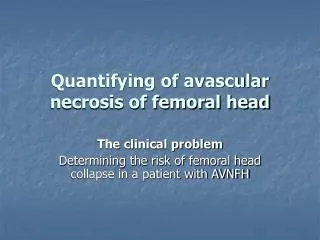
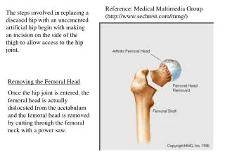
Clinical Evaluation of A Novel Reconstruction Device for Osteonecrotic Femoral Head. Ching-Chuan Jiang M.D., Ph.D., M.B.A. Arthropathy Research Laboratory. Osteonecrosis of Femoral Head.

E N D
Clinical Evaluation of A Novel Reconstruction Device for Osteonecrotic Femoral Head Ching-Chuan Jiang M.D., Ph.D., M.B.A. Arthropathy Research Laboratory
Osteonecrosis of Femoral Head • Out of all total hip replacements in the US and Europe, 10% are caused by osteonecrosis with approximately 300,000 to 600,000 casesof osteonecrosis of femoral head in the US every year. • Asian area has a higher rate as 50%oftotal hip replacements are due to osteonecrosis.
MRI Staging of Osteonecrotic Femoral Head Stage I Normal radiograph Stage II Femoral head Lucency/sclerosis on radiograph Stage III Subchondral collapse without femoral head flattening, “crescent sign“ Stage IV Subchondral collapse, femoral head flattening, normal joint space Stage V Flattening with joint space narrowing, acetabular changes, or both Stage VI Advanced degenerative changes, secondary osteoarthritis
Knee and extremity joints cartilage repair technologies • (Biphasic Cartilage Repair Implant,BiCRI) have been transferred to Exactech in 2008. Progresses and results were reported in the ongoing clinical trial. • Applications on indications related to hip joint, craniofacial and spine are still retained in NTUH/ITRI and not yet being transferred. Clinical Trial: Efficacy Evaluation of ONFH Reconstruction Device
Current Patent Status • P56980010US, temporary application no.:12/830,536. Patent name: Bone Repairing Kit and Method for Bone Repair • P56980010TW, Patent no.:I413504 Patent name:骨修復套組
Patents for Biphasic Matrix • Process for producing porous polymer materials, US Pat. No • 6,436,426 • Method for tissue culture in vitro, US Pat. No 6,884,621 • Process for producing porous polymer materials, US Pat. No • 6,824,716 • Method and carrier for culturing multi-layer in vitro, I255852 • Tissue pulverizer, I 255738 • Multi-layered matrix, method of tissue repair using the same, • and multi-layered implant prepared thereof, US Pat. Revised. • Filtration and collection device for tissue and cells, US Pat. • Filing. Seven patents have been issued.
Subjects selection Preoperative evaluation Standard Operating Procedure Control (core decompression) Experimental (ONFH Repair Device) Postoperative evaluation and data analysis Planned to operate 20 cases: Including 10 in control group (core decompression) and 10 inexperimental group (ONFH device)
Recruitment status: • I.Inclusion criteria: • No allergies. • No critical illness. • 20 – 70 years old. • Shape of the femoral head is generally preserved and the lesion diameter is less than 1.8 cm on radiographs, CT and MRI scans. • MRI Staging III lesion.
II. Exclusion criteria: • Prior surgical treatment of the lesion. • Pregnancy. • Extensive degenerative hip arthritis. • Osteoporosis in femoral head and neck. • Rheumatoid arthritis and other inflammatory arthritis of hip. • Hip stiffness, defined as: • Flexion < 20 degrees. • Internal rotation < 90 degrees. • Lesions: MRI Staging IV, V and VI.
III. The evaluation methods: • Assess the subchondral bone and articular cartilage by MRI.
IV. Experiment time schedule: Evaluation methods Post surgery Follow up (weeks)
V. Data collection and Statistical analysis: • DATA collection between pre- and post-surgery: • Hip range of motion • Harris Hip Score • WOMAC (The Western Ontario and McMaster Universities) Osteoarthritis index • VAS(Visual Analogue Scale) • Statistical analysis: • One-tailed Student’s t-test: Hip range of motion, VAS • Wilcoxon Rank Sum test: Harris Hip Score, WOMAC
Pre-Clinical Animal Study Development of implantable scaffold and surgical instrument completed • Cannulated reamer • Ejector • Holder • Trimmer • Resorbable Bone Substitute Hip joint reconstruction surgical instrument and implantable scaffold Design and development of surgical procedure and 8 porcine large animalimplantation tests completed; articular cartilage and bone at femoral head can be reconstructed and repaired.
Patient profile Case # 01 (Op: 2013.08.29) • 37 y/o female • Wt / Ht: 72kg / 158.2cm • Diagnosis: Osteonecrosis of femoral head, right hip • Complaint: right hip pain for 8 months • History: steroid used 2 years for Vasculitis • OP Method: A novel reconstruction device
Case# 01 X-Ray Pre-OP Post-OP 6weeks Post-OP 3months
Patient profile Case # 02 (Op: 2013.10.24) • 56 y/o female • Wt / Ht: 57.8kg / 163.6cm • Diagnosis: Osteonecrosis, left femoral head • Complaint: Left hip pain for 6 months • History: HB carrier, liver abscess • OP Method: A novel reconstruction device
Case# 02 X-Ray Post-OP 6 weeks Pre-OP
Case # 02 Tissue and Histology Section A Section B 40x 40x 1 cm 100x 400x 400x
Patient profile Case # 03 (Op:2013.12.05) • 29 y/o female • Wt / Ht: 53.6kg / 162.3cm • Diagnosis: Osteonecrosis, left femoral head • Complaint: 1.Low back pain with frequent soreness 2.decreased range motion and pain at left hip for 2 years • History: Not contributory • OP Method: Core decompression
Case# 03 X-Ray Pre-OP Immediate Post-OP
Preliminary Conclusions • Two patients of the experiment group (osteonecrotic • device) and one patient of the control group (core • decompression) were performed uneventfully. • No perioperative complications occurred. • Immediate pain relieve in three patients of both groups • were observed. • The experimental procedure can provide a minimal • invasive endoscopic approach for direct visual examination • to the lesion site. It also open a venue for further • therapeutic managements to the osteonecrotic femoral • head.
Acknowledgement • National Taiwan University College of Medicine: Hongsen Chiang, Jyy-Jih Tsai, Chang-Hsun Hsieh, Chia-Jung Tsai, Sy-Chi Chen, Ya-Pin Wu, Rung-Shan Lee • Industrial Technology Research Institute: Chun-Jen Liao, Wen-Hsiang Chang, Huang-Chi Chen, Huang-Chi Chen, Ming-Chia Yang, Wei-Ju Liao , Yun-Han Lin , Chia-Chi Ho, Wei-Hong Chang, Ping-Chung Chang, Fang-Jie Jang, Chi-Hsiang Liao • Intai Technology Corp • Financial Support: NRPB,ITRI,NTUH