Download
1 / 19
380 likes | 2.18k Views
Lecture 13: Mechanism of Chymotrypsin. Chemical Mechanism of Chymotrypsin. Chymotrypsin. Chymotrypsin is a digestive protease involved in breakdown of proteins and peptides so that their amino acids can be used. It is synthesized in the pancreas of mammals and released into

E N D
Lecture 13:Mechanism of Chymotrypsin Chemical Mechanism of Chymotrypsin
Chymotrypsin Chymotrypsin is a digestive protease involved in breakdown of proteins and peptides so that their amino acids can be used. It is synthesized in the pancreas of mammals and released into the digestive tract. When first synthesized it is as a single polypeptide chain in an inactive form, chymotrypsinogen, which must be activated before the enzyme can fulfill its role. Activation of chymotrypsin is achieved by “clips” in its polypeptide chain, so active chymotrypsin consists of three distinct chains. These remain bound together in a single domain, covalently held together by disulfide bonds. The activity of chymotrypsin is regulated by controlling when the “clips” are made.
Amide + H2O Acid + Amine DG uncat DG Net Reaction: Overall DG of hydrolysis is negative (favorable). Uncatalysed pathway cat Reactants DG’ Enzyme-catalysed pathway Products
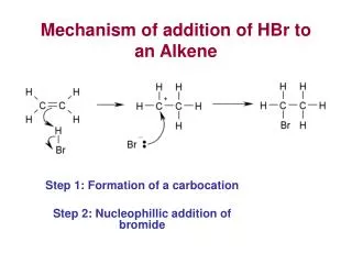
Overview: S PN PC (Bond to be cleaved.) Substrate (S) binds. Phase 1: Enzyme creates nucleophile from serine side-chain. Nucleophile attacks substrate. Covalent intermediate is formed with second product ( PN ) bonded to serine, and first product ( PC ) is released. Phase 2: Enzyme creates a nucleophile from a water molecule. Nucleophile attacks covalent intermediate, breaking covalent bond to serine. Second product ( PN ) is released. PN S PC E ES EPN E-PN PC E-PN Substrate Binding Chemical Rearrangement Product #1 Released Chemical Rearrangement Product #2 Released
DG E+S ES E-PNPC E-PN + PC EPN + PC E+PN +PC cat EPN Reactants DG’ ES E-PNPC Products
Clues about mechanism: Burst phase indicates a covalent intermediate is formed. Kinetics experiments are used to figure out how many steps there are in a reaction mechanism and how long each step takes. Chemical labeling with DIPF finds one particular serine residue (out of 28) that is extremely reactive. Chemical labeling experiments are used to figure out which residues are responsible for important steps in a reaction mechanism. Crystal structure analysis reveals a catalytic triad, a group of 3 side-chains which are responsible for the peculiar reactivity of this serine. Determination of the crystal structure of an enzyme provides a detailed description of the three-dimensional arrangement of the molecule and in particular of the active site.
Chymotrypsin Kinetics (remains covalently bound) (released immediately) Very early in reaction, p-nitrophenolate is released giving rise to the burst phase. Subsequent reactants must wait for an active site to become available through release of an intermediate, giving rise to the steady-state phase. Km = 20 mM kcat = 77 s-1
Labelling of Serine 195 Inactivates Chymotrypsin DIPF, an irreversible inhibitor, is a group-specific reagent for serine residues. It forms a covalent adduct on Serine 195, which renders the enzyme inactive. Only Ser 195, out of 28 serines in chymotrypsin, is labelled, suggesting it is both especially reactive and that this reactivity is necessary for catalysis.
Structure of Chymotrypsin Globular single-domain protein. Originally synthesized as a 245 residue protein, chymotrypsinogen. Dipeptides 14-15 and 147-148 are clipped out, tranforming the protein into active chymotrypsin. Therefore it has 3 chains (red, blue, green) but these are covalently linked by disulfide bridges. The reactive serine 195 is located in a cleft on the molecule, the active site. Ser 195 is adjacent to His 57 and Asp 102 which are responsible for its reactivity.
Substrate (S) binds. Enzyme creates nucleophile from serine side-chain. Nucleophile attacks substrate. Acyl-enzyme is formed with second product ( PN ) bonded to serine, and first product ( PC ) is released. Enzyme creates a nucleophile from a water molecule. Nucleophile attacks acyl linkage, breaking covalent bond to serine. Second product ( PN ) is released. Phase 1: Phase 2:
Creation of Nucleophile A nucleophile is a highly reactive, electron-rich group. In chymotrypsin, serine 195 is converted into an alkoxide ion, a powerful nucleophile, through removal of its hydroxyl proton. This difficult task is accomplished by the charge relay system between Asp 102, His 57, and Ser 195, which comprise the catalytic triad. His 57 can alternately accept or donate protons, while stabilized by Asp 102. (This is a good example of a general base in catalysis.) The charges are stabilized by electrostatic effects.
Nucleophilic Attack The carbonyl carbon on the substrate has 3 bonds and so is a trigonal atom. The alkoxide ion attacks the carbonyl carbon, forming a tetrahedral intermediate with 4 bond to that carbon. The former carbonyl oxygen is converted into a negatively charged group, the oxyanion, which is stabilized by by an arrangement of partial positive charges nearby in the oxyanion hole. (an electrostatic effect)
Formation of Acyl-enzyme The tetrahedral intermediate breaks down when the histidine donates a proton and creates a new amino group on the terminus of the first product ( PC ), which is released. (His 57 is acting as general acid.) The remainder of the substrate remains attached to the enzyme through an ester linkage to Serine 195. (Covalent catalysis.) Acyl group
Creation and Use of New Nucleophile Another nucleophile is created by the enzyme, using His 57 to withdraw a proton from a water molecule to form a hydroxide ion. (another example of general base catalysis) This nucleophile attacks the acyl carbon forming a second tetrahedral intermediate, which is again stabilized by the oxyanion hole. (Another example of the electrostatic effect.)
De-acylation Step The tetrahedral intermediate breaks down when His 57 donates a proton to serine 195, displacing the acyl group and regenerating the serine hydroxyl group. The second product ( PN ) is released, concluding the reaction.
Same Chemistry, Different Enzymes Other unrelated classes of proteases, whose sequences and structures are unrelated to those of chymotrypsin, nevertheless have the same spatial arrangement of the His-Asp-Ser catalytic triad. The same catalytic method appears to have arisen independently at least three times in nature- an example of convergent evolution. Subtilisin Carboxypeptidase A
Summary: Chymotrypsin is a protease and its activity is regulated by controlled cleavage of its backbone. Its chemical mechanism proceeds in two stages: 1. Nucleophilic attack on substrate by Ser 195 to form acyl-enzyme complex followed by 2. Deacylation though nucleophilic attack by water on the acyl intermediate. Key Concepts: Meaning of burst phase and labelling of Serine 195 Catalytic triad: Roles of His 57, Asp 102, and Ser 195 in mechanism Occurrences of acid-base catalysis and covalent catalysis in mechanism