Download
1 / 59
610 likes | 1.34k Views
Diabetes Mellitus and Disorders of Glucose Homeostasis. Rosen’s Chapter 124 December 21, 2006 Presented by: Dr. D’Isa-Smith Prepared by: Michael Savino, DO (PGY-2). Normal Physiology. Normal glucose range: 60-150 mg/dL

E N D
Diabetes Mellitus and Disorders of Glucose Homeostasis Rosen’s Chapter 124 December 21, 2006 Presented by: Dr. D’Isa-Smith Prepared by: Michael Savino, DO (PGY-2)
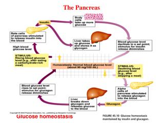
Normal Physiology • Normal glucose range: 60-150 mg/dL • Normal plasma glucose levels are critical to survival, because glucose is main fuel for CNS • CNS does not synthesize glucose and only stores a few minutes supply of glucose. • Brief hypoglycemia can cause profound brain dysfunction • Prolonged severe hypoglycemia can cause cellular death
Glucose • Derived from 3 sources: • 1. intestinal absorption • 2. glycogenolysis – glycogen breakdown • 3. gluconeogenesis – glucose formed from precursors such as lactate, pyruvate, amino acids, glycerol • After glucose ingestion, plasma levels rise and endogenous production is suppressed.
Insulin • b cells of the pancreas detect elevated glucose levels triggering release of insulin into the hepatic portal circulation • Major anabolic hormone in diabetic disorder • Stimulates glucose uptake, storage, and use by other insulin-sensitive tissues (fat, muscle) • Half-life of insulin is about 3-10 minutes • Metabolized through the liver and kidney
Liver and Kidney • Liver and kidney contain glucose-6-phosphatase – enzyme necessary for the release of glucose into the circulation • The liver is the sole source of endogenous glucose production in normal conditions • The kidney undergoes gluconeogenesis under prolonged starvation
Hepatocytes do not require insulin for glucose transport across cell membrane • But, insulin augments hepatocyte glucose uptake and storage for energy • Insulin inhibits hepatic gluconeogenesis and glycogenolysis
Muscle cells • Can store and use glucose via glycolysis • In muscle: glucose pyruvate • Pyruvate lactate or alanine transported to liver precursor for gluconeogenesis Fasting conditions: i glucose uptake – use fatty acids as energy, mobilize amino acids to liver for energy.
Counterregulatory hormones • Glucagon • The major catabolic agent that increases blood glucose • a cells of pancreas • Released in response to hypoglycemia, stress, trauma, infection, starvation. • Decreases glycoloysis, increases gluconeogenesis • Increases ketone production in liver • Epinephrine – h hepatic glucose production and limits glucose use through a and b adrenergic mechanisms • acts directly: glycogenolysis, gluconeogenesis • Norepinephrine – similar to epinephrine • Growth hormone and cortisol – initially i glucose, but long-term h glucose
Types of Diabetes • Type 1 Diabetes Mellitus • Type 2 Diabetes Mellitus • Gestational Diabetes • Impaired Glucose Tolerance
Type 1 DM • Failure to produce insulin. Tendency to ketosis • Parenteral insulin required to sustain life • Autoimmune destruction of Beta cells of pancreas • Strong association with HLA
Type 1 DM • Typical patient is lean, younger than 40, prone to ketosis. • Plasma insulin levels are low or absent. • Glucagon levels are high, but suppressible with insulin • Symptoms of polydipsia, polyuria, polyphagia, and wt. loss develop rapidly • Complications incl: DKA, retinopathy, nephropathy, neuropathy, foot ulcers, severe infections
Type 2 DM • Typical patient is middle aged or older, overweight, normal to high insulin levels. • Impaired insulin function related to poor insulin production, failure of insulin to reach the site of action, or failure of end organ response to insulin • Symptoms begin more gradually than in Type 1
Type 2 DM - Subgroups • Most are obese, but 20% are not • Nonobese Type 2 patients present more like Type 1 • Young persons with mature-onset diabetes
Type 2 DM • Symptoms come on gradually • Diagnosis usually made by elevated blood glucose on routine lab work • Blood glucose levels controlled by diet, oral hypoglycemics, or insulin. • Decompensation usually leads to hyperosmolar nonketotic coma rather than ketosis.
Gestational Diabetes • Characterized by abnormal oral glucose tolerance test (OGTT). • During pregnancy • Reverts to normal in postpartum period or remains abnormal • Clinical pathogenesis similar to Type 2 • Clinical presentation usually nonketotic hyperglycemia during pregnancy
Impaired glucose toleranceImpaired Fasting Glucose • Plasma glucose levels between normal and diabetic and who are at increased risk for development of diabetes • Pathogenesis related to insulin resistance • Presentations: nonketotic hyperglycemia, insulin resistance, hyperinsulinism, often obesity • Less complications than diabetes
Diagnostic Strategies Diagnosis made by: • Random plasma glucose > 200 mg/dL • or Fasting glucose > 140 mg/dL • or 2 hr postload OGTT • HbA1c – high glucose binds to Hb b chain. Half-life of RBC’s allows index of [glucose] for prior 6-8 weeks (normal 4-6%) • Glucose dipstick tests use glucose oxidase • Ketone dipstick tests use nitroprusside rxn.
Dipstick Blood Glucose (Accucheck) • Generally more accurate than urine dip • Hematocrits <30% or >55% cause unduly high or low readings, respectively
Hypoglycemia • Common problem in Type 1 diabetics • 9 – 120 episodes per 100 patient-years • Severe hypoglycemia associated with blood sugar below 40-50 mg/dL and impaired cognitive function • Hypoglycemia unawareness – a dangerous complication of Type 1. Pts become unarousable without warning • Somogyi phenomenon
Hypoglycemia - symptoms • Blood glucose level below 40-50 mg/dL • Rate at which glucose decreases, age, gender, overall health, and previous hypoglycemic reactions all contribute to symptom severity • S/Sx caused by excessive epinephrine secretion and CNS dysfunction: • Sweating • Nervousness, tremor • Tachycardia • Bizarre behavior, confusion • Seizures • Coma
Hypoglycemia - treatment • 1. Suspect hypoglycemia • Check serum glucose; if strong suspicion treat before results available • 2. Correct serum glucose • If awake, cooperative: PO intake • If unable to take PO: 25-75 g glucose as D50W (1-3 amps) IV • Children: 0.5-1 g/kg glucose as D25W IV • Neonates: 0.5-1 g/kg glucose as D10W IV • If unable to get IV access: 1-2 mg glucagon IM or SC; may repeat q20 min • Glucagon – onset of action 10-20 min, peaks at 30-60 min • Ineffective in alcohol-induced hypoglycemia b/c lack of glycogen
Hypoglycemia - Management • ABC’s • Aspiration, seizure precautions • If ETOH suspected, give thiamine • D50W should not be used in infants or young children because venous sclerosis causes rebound hypoglycemia • Oral hypoglycemics (chlorpropamide) – can cause prolonged hypoglycemia. Should be admitted for observation • May require constant infusion of D10W
Hypoglycemia • Non-diabetic patients • Most common cause of postprandial hypoglycemia is alimentary hyperinsulinism (s/p gastrectomy, gastrojejunostomy, vagotomy, pyloroplasty) • Fasting hypoglycemia – inadequate glucose production (hormone deficiencies, enzyme and substrate defects, severe liver disease)
Hyperglycemia:Diabetic Ketoacidosis • Syndrome in which insulin deficiency and glucagon excess produce: • Hyperglycemia • Dehydration • Acidosis • Electrolyte imbalance • DKA is typically characterized by: • Hyperglycemia over 300 mg/dL, • Low bicarbonate (<15 mEq/L), and • Acidosis (pH <7.30) with ketonemia and ketonuria
Etiology of DKA • Almost always in Type 1 Diabetics • Non-compliance with insulin • Stress: (Physical or emotional) despite insulin use • Myocardial infarction • Infection/ Sepsis • Gastrointestinal bleeding • 25% of all episodes of DKA occur in undiagnosed patients.
DKA: History and Physical • Polydipsia, polyuria, polyphagia, visual blurring, weakness, wt loss, N/V, abd pain • May have altered mental status • Kussmaul respirations • Odor of acetone (sweet) on breath • Signs of dehydration • Tachycardia • Orthostatic changes
Pathophysiology DKA • Markedly elevated glucose levels spill over into the urine, drawing water, sodium, potassium, magnesium, calcium, phosphorus into the urine. • This combined with vomiting contribute to dehydration experienced in DKA • Exocrine pancreas dysfunction produces malabsorption, further limiting body’s intake of fluid and electrolytes.
Falsely elevated Elyte levels • 95% of DKA patients: • Na = normal or low • K = very low (5-7 mEq/L) • Mg = very low • Phos = very low (3 mEq/L) • Because of dehydration and acidosis, however, these lab values are reported as high!
Ketosis/Acidosis • Adipose tissue fails to clear the circulation of lipids. Insulin deficiency results in activation of hormone-sensitive lipase increasing free fatty acid [FFA] levels. Overload of FFA’s on the liver oxidizes them to acetoacetate and Beta-hydroxybuterate. • Result is oxidation of FFA’s to ketones instead of reesterification to triglycerides • The body while increasing ketone production, utilizes less ketones in peripheral tissues leading to ketoacidosis.
Ketoacidosis • Glucagon levels are 4-5x higher in DKA and is the most influential ketogenic hormone. • Glucagon inhibits malonyl coenzyme A and inhibits glycolysis
The counterregulatory hormones: Epinephrine, norepinephrine, cortisol, growth hormone, dopamine, and thyroxin enhance ketogenesis indirectly by stimulating lipolysis. • Propranolol and metyrapone can block the effect of counterregulatory hormones. They have been used to prevent recurrent episodes in known DKA patients.
Acidosis in clinical presentation • Acidotic patient attempts to increase lung ventilation and rid the body of excess acid with Kussmaul’s respiration. Bicarbonate is used up in the process. • Current evidence suggests that acidosis compounds the effects of ketosis and hyperosmolality to depress mental status directly.
Pathophysiology of DKA Adapted from figure 124-1, p. 1963
Laboratory Tests • Allow confirmation of diagnosis • [ serum and urine glucose (usually greater than 350, but up to 18% of patients may have euglycemic DKA) • [ Electrolytes • [ ABG/venous pH (w/ K+ if available) • Obtain EKG immediately
Metabolic acidosis • Metabolic acidosis with elevated anion gap is secondary to elevated plasma levels of acetoacetate and b-hydroxybutyrate. Also contributed by lactate, FFA’s, phosphates, volume depletion
Other tests • CBC w/ differential • BMP – elevated BUN/Cr suggest dehydration. • Mag, calcium, amylase, ketone, and lactate levels • U/A – rule out infection/renal dz
Sodium • Serum sodium value often misleading! • Sodium is often low in presence of dehydration because affected by: • Hyperglycemia • Hypertriglyceridemia • Salt-poor fluid intake • Insensible losses • Marked hyperglycemia – water flows from cells into vessels to decrease osmolar gradient, causing dilutional hyponatremia • Correction: Na + (Gluc – 100) * 1.6 / 100 • For every increase of 100 mg/dL glucose, the serum sodium decreases by 1.6
Hypertriglyceridemia • Common in DKA • Impaired lipoprotein lipase activity and hepatic overproduction of VLDL
Acidosis • Acidosis and hyperosmolarity by high glucose levels shift potassium, magnesium, and phosphorous from intracellular to extracellular space. • Dehydration produces hemoconcentration, which contributes to normal-high initial serum potassium, mag, and phos
Calculate Correction for potassium • Correction for the effects of acidosis on serum potassium: • Subtract 0.6 mEq/L from lab K+ for every 0.1 decrease in pH on ABG’s • Ex: if K+ is reported as 5 mEq/L and the pH is 6.94, the corrected K+ = 2 mEq/L
Management of DKA • Consider intubation in vomiting decompensated patient for airway protection • Once intubated, hyperventilation should be maintained to prevent worsening acidosis • Hypovolemic shock: requires aggressive fluid resuscitation with 0.9% NSS, rather than pressors • Consider other causes of shock: MI, sepsis • Diagnosis: Hyperglycemia, ketosis, acidosis • Fluids, electrolytes, insulin therapy begins.
Summary of treatment for DKA Identify DKA: glucose, electrolytes, ketones, ABG. CBC, U/A, CXR, EKG. Support ABC. • 1. Rehydrate: 1-2 L NSS over 1-3 hours • Children: 20 mL/kg NSS over first hr, then follow w/ 0.45% NSS • 2. Insulin – bolus 0.1 U/kg regular IV • Maintenance: 0.1 U/kg/hr regular IV • Change to D5W/0.45%NS when glucose <300 mg/dL • 3. Correct electrolytes. • Na – 0.9% NSS and 0.45% • K – add 20-40 mEq KCl to each liter. Ensure good renal fxn • Phos – usually not necessary to replenish • Mg – 1-2 g MgSO4
4. Correct acidosis – add 44-88 mEq/L bicarb to 1st liter of IV fluids if pH < 7.0. Correct to a pH of 7.1 • Correct underlying precipitant • Monitor VS, I & O’s, serum glucose, and electrolytes • Admit to ICU
Insulin • Historically, high dosages of insulin were used, but resulted in hypoglycemia and hypokalemia • Now, low-dose insulin therapy with aggressive fluid therapy is used, more gradual decrease in blood glucose levels, while decreasing risk of hypokalemia
Insulin • May start with bolus of 10 units regular insulin • Or infuse regular insulin at a rate of 0.1 U/kg/hr up to 5-10 U/hr, mixed with IV fluids. • In children, dosing is 0.1 U/kg. Reduction of plasma glucose should be more gradual because of greater risk of developing cerebral edema.
Half-life of regular insulin is 3 – 10 minutes. • Therefore, it should be infused, rather than given as repeated boluses. • When blood glucose has dropped to 250-300 mg/dL, then start D5W/0.45% NS to prevent iatrogenic hypoglycemia and cerebral edema
DKA Glucose >350 Sodium ~ low 130s Potassium ~ 4.5-6.0 Bicarbonate < 10 BUN ~25-50 Serum ketones present HHNC Glucose >700 Sodium ~ 140s Potassium ~ 5 Bicarbonate > 15 BUN > 50 Serum ketones absent DKA vs. HHNC
Hyperglycemic Hyperosmolar Nonketotic Coma • Acute diabetic decomposition • Results from severe dehydration that results from sustained hyperglycemic diuresis, in which patient is unable to drink enough fluids to sustain hydration • Characterized by: Hyperglycemia, hyperosmolarity, dehydration • Absence of ketoacidosis is unknown, but FFA levels are lower than in DKA, thus less substrates to form ketones. Most likely because still producing tiny amount of insulin required to block ketogenesis
More common in elderly with Type 2, but has been reported in children with Type 1 • May occur in pts who are not diabetic: after burns, parenteral hyperalimentation, peritoneal dialysis, or hemodialysis • Clinically: signs of dehydration and CNS findings predominate • Most common associated diseases: CRI, gm – pneumonia/sepsis, GI bleeding • On average, the HHNC patient has 24% or 9L fluid fluid deficit
Treatment of HHNC • Identify HHNC • Rehydrate: 2-3 L NSS over first few hours. Correct ½ fluid deficit in first 8 hours, remainder over remaining 24 hrs • Insulin – bolus 0.05-0.1 U/kg regular IV • Maintenance: 0.05-0.1 U/kg/hr regular IV • Change to D5W/0.45%NS when glucose <300 mg/dL • . Correct electrolytes. • Na – 0.9% NSS and 0.45% • K – add 20-40 mEq KCl to each liter. Ensure good renal fxn • Phos – usually not necessary to replenish • Mg – 1-2 g MgSO4